. 2023 Aug 31;15(9):2263.
doi: 10.3390/pharmaceutics15092263.

A Species-Specific Anti-Human P2X7 Monoclonal Antibody Reduces Graft-versus-Host Disease in Humanised Mice

Affiliations

A Species-Specific Anti-Human P2X7 Monoclonal Antibody Reduces Graft-versus-Host Disease in Humanised Mice

Amal Elhage et al. Pharmaceutics. .

Abstract

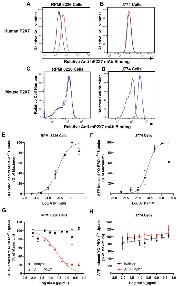
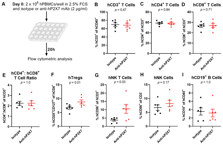
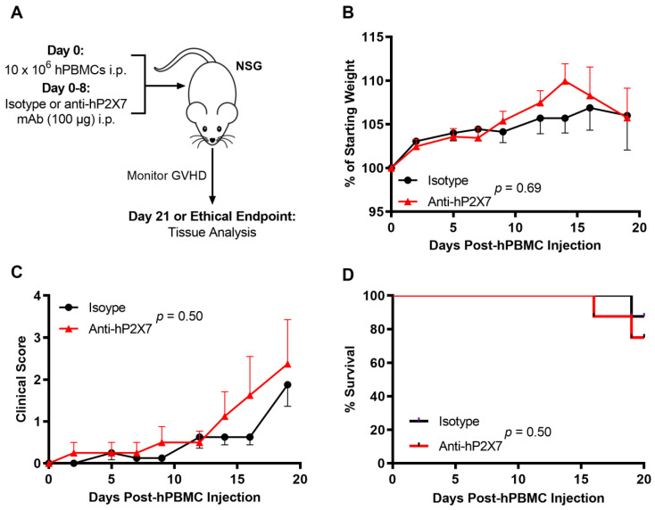
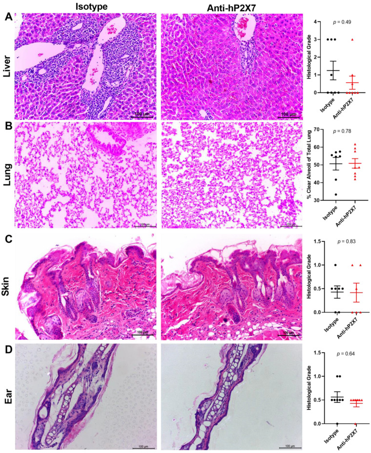
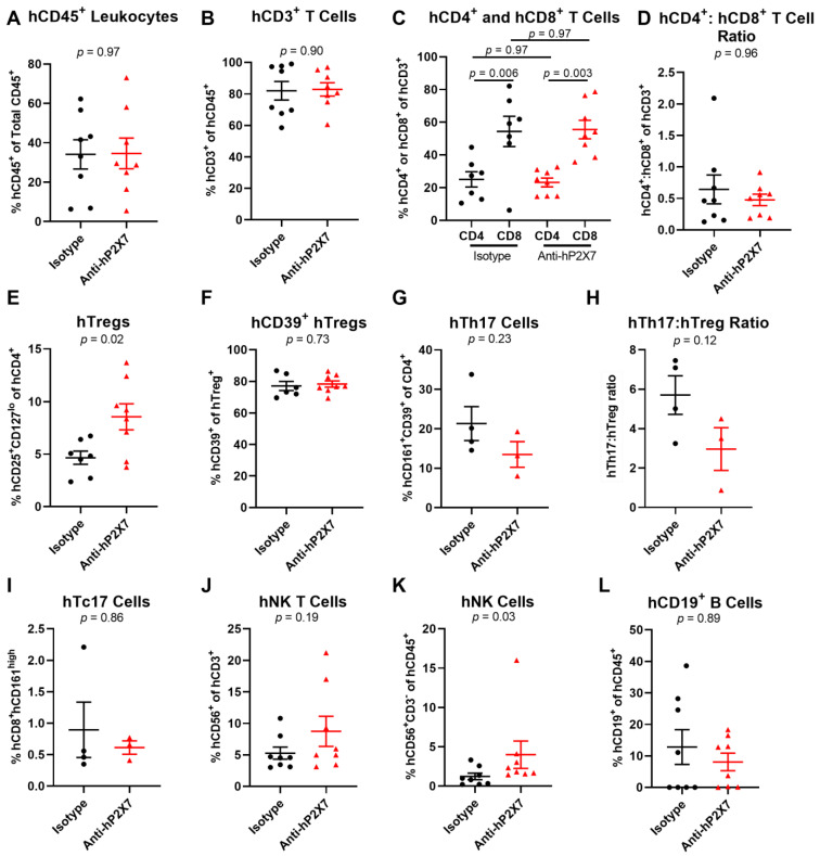
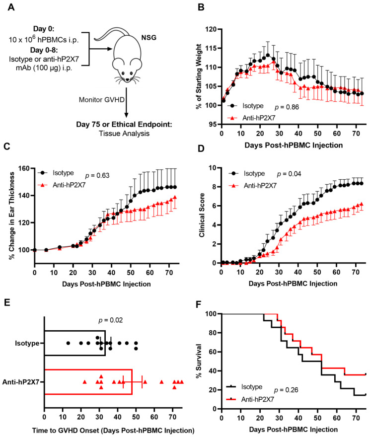
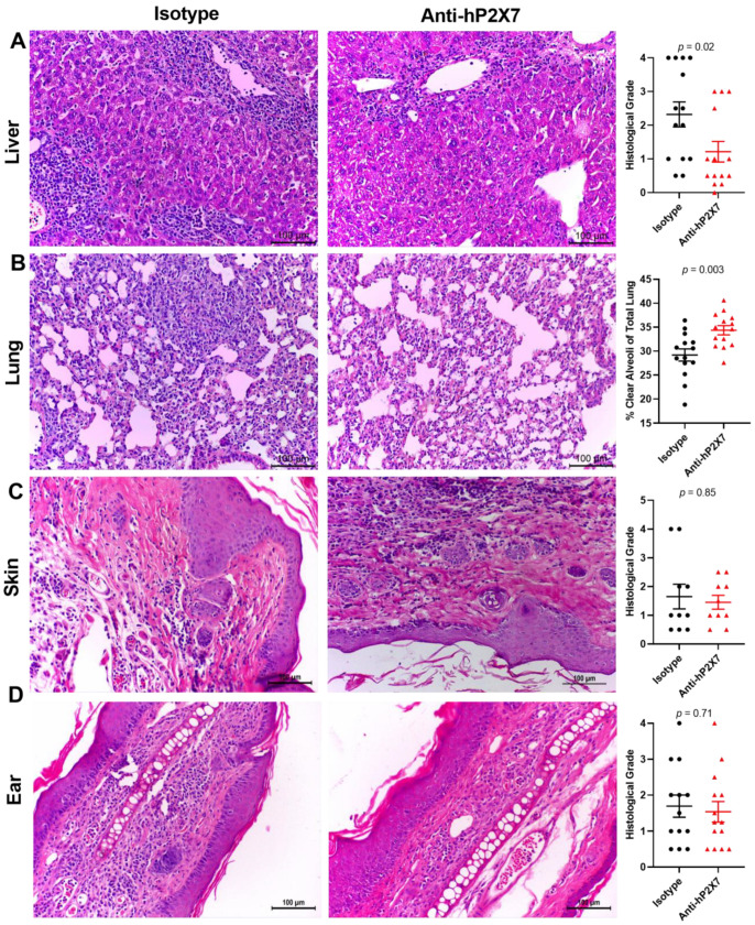
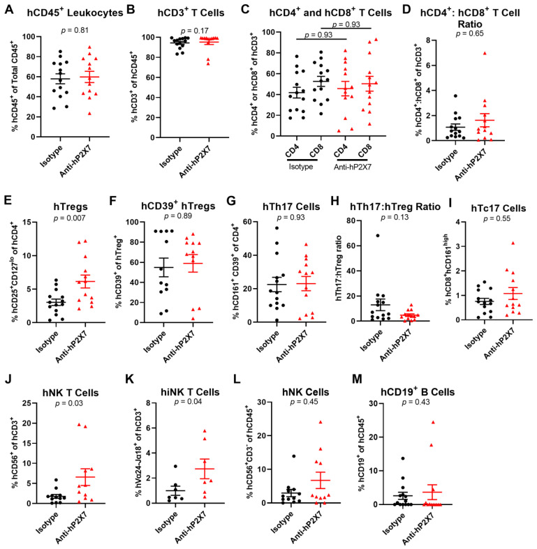
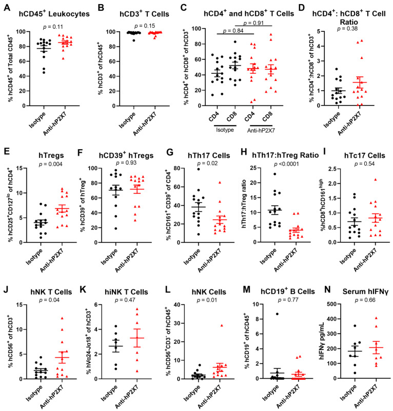
Graft-versus-host disease (GVHD) is a T cell-mediated inflammatory disorder that arises from allogeneic haematopoietic stem cell transplantation and is often fatal. The P2X7 receptor is an extracellular adenosine 5'-triphosphate-gated cation channel expressed on immune cells. Blockade of this receptor with small molecule inhibitors impairs GVHD in a humanised mouse model. A species-specific blocking monoclonal antibody (mAb) (clone L4) for human P2X7 is available, affording the opportunity to determine whether donor (human) P2X7 contributes to the development of GVHD in humanised mice. Using flow cytometric assays of human RPMI 8266 and murine J774 cells, this study confirmed that this mAb bound and impaired human P2X7. Furthermore, this mAb prevented the loss of human regulatory T cells (hTregs) and natural killer (hNK) T cells in vitro. NOD-scid IL2Rγnull mice were injected with 10 × 106 human peripheral blood mononuclear cells (Day 0) and an anti-hP2X7 or control mAb (100 μg i.p. per mouse, Days 0, 2, 4, 6, and 8). The anti-hP2X7 mAb increased hTregs and hNK cells at Day 21. Moreover, anti-hP2X7 mAb-treatment reduced clinical and histological GVHD in the liver and lung compared to the control treatment at disease endpoint. hTregs, hNK, and hNK T cell proportions were increased, and human T helper 17 cell proportions were decreased at endpoint. These studies indicate that blockade of human (donor) P2X7 reduces GVHD development in humanised mice, providing the first direct evidence of a role for donor P2X7 in GVHD.

Keywords: P2RX7; P2X7; T helper 17 cells; biologic; natural killer T cells; natural killer cells; purinergic signalling; regulatory T cells; therapeutic antibody; xenogeneic graft-versus-host disease.


Conflict of interest statement

The authors declare no conflict of interest. The funders had no role in the design of the study; in the collection, analyses, or interpretation of data; in the writing of the manuscript; or in the decision to publish the results.

Figures

Figure 1
Figure 1
The anti-hP2X7 mAb binds and blocks human but not mouse P2X7. (A,C) Human RPMI 8226 and (B,D) mouse J774 cells were incubated with (A,B) DyLight488 conjugated anti-hP2X7 (red line) or isotype control (black line) mAb or (C,D) PE-conjugated anti-mP2X7 (blue line) or isotype control (black line) mAb and analysed by flow cytometry. (E) Human RPMI 8226 (n = 4) and (F) mouse J774 cells (n = 4) in LDM containing 1 µM YO-PRO-12+ were incubated with 0.02 to 2 mM of ATP at 37 °C for 5 min. (G) Human RPMI 8226 (n = 3) and (H) mouse J774 cells (n = 3) in LDM were pre-incubated with 0.01 µg/mL to 5 µg/mL of the anti-hP2X7 or isotype control mAb at 37 °C for 10 min. Cells were then incubated with 1 µM of YO-PRO-12+ in the absence or presence of 0.3 mM and 0.26 mM ATP for human RPMI 8226 and mouse J774 cells, respectively, at 37 °C for a further 5 min. (EH) Incubations were terminated by the addition of ice-cold medium containing MgCl2 and centrifugation. YO-PRO-12+ uptake was then analysed by flow cytometry and data normalised to the maximum ATP response in each experiment. (EH) Results are the mean ± SEM.
Figure 2
Figure 2
The anti-hP2X7 mAb prevents the loss of hTregs and hNK T cells in vitro. (A) Illustration of the in vitro cultures. (BI) hPBMCs (n = 6 donors) were cultured overnight (20 h) in RPMI-1640 medium containing 2.5% FCS in the presence of 2 µg/mL of the isotype control or anti-hP2X7 mAb. The proportions of (B) hCD3+ T cells, (C) hCD4+ and (D) hCD8+ T cell subsets, (F) hCD4+hCD25+hCD127lo hTregs, (G) hCD56+ hCD3+ hNK T cells, (H) hCD56+ hCD3 hNK cells, or (I) CD19+ B cells were determined by flow cytometry. (E) The ratio of hCD4+ to hCD8+ T cells was calculated from (C,D). (BI) Data represented as mean ± SEM, symbols represent individual donors. Significance was assessed by the paired Student’s t-test, with p values as shown.
Figure 3
Figure 3
The anti-hP2X7 mAb did not affect early clinical GVHD development in humanised mice over 21 days. (A) Illustration of a humanised mouse model of GVHD. NSG mice were injected with 10 × 106 hPBMCs (n = 2 donors) at Day 0 followed by 100 μg of the isotype control or anti-hP2X7 mAb every second day from Days 0–8 (n = 8 mice for each group). Mice were monitored thrice weekly over 21 days for (B) weight loss, (C) clinical score or (D) survival. (B,C) Data represented as mean ± SEM. Significance was assessed by the (B,C) two-way ANOVA with a Bonferroni post-test or (D) Mantel–Cox log-rank test for survival, with p values as shown.
Figure 4
Figure 4
The anti-hP2X7 mAb does not affect histological GVHD in the liver, lung, skin, and ear of humanised mice at Day 21. The (A) liver, (B) lung, (C) skin, and (D) ear from isotype control (n = 7–8) or anti-hP2X7 (n = 6–8) mAb-treated mice at Day 21 (or humane endpoint) were sectioned (3–5 μm), stained, and graded based on evidence of histological GVHD. (A,C,D) Liver, skin, and ear were measured using a grading system. (B) Histological GVHD in the lung was measured as the percentage of clear alveoli space of total lung area. Images represent at least six mice per treatment group. (AD) Scale bars represent 100 µm and data represented as the mean ± SEM. Symbols represent individual mice. Significance was assessed by the Mann–Whitney test, with p values as shown.
Figure 5
Figure 5
The anti-hP2X7 mAb increases proportions of splenic hTregs and hNK cells in humanised mice at Day 21. (AL) Spleens from mice treated with the isotype control (n = 8) or anti-hP2X7 (n = 8) mAb were collected at Day 21 (or humane endpoint) and immune cell subsets were analysed by flow cytometry. Proportions of (A) hCD45+ leukocytes were first identified before determining the proportions of (B) hCD3+ T cells, (C) hCD4+ and hCD8+ T cells, (E) hCD4+hCD25+hCD127lo hTregs, (F) hCD39+ hTregs, (G) hCD4+hCD161+hCD39+ hTh17 cells, (I) hCD8+hCD161high Tc17 cells, (J) hCD3+hCD56+ hNK T cells, (K) hCD3hCD56+ hNK cells, and (L) hCD19+ B cells. (D) The hCD4+:hCD8+ T cell ratio was calculated from (C). (H) The hTh17:hTreg ratio was calculated from (E,G). (AL) Data represented as the mean ± SEM. Symbols represent individual mice. Significance was assessed by the (A,B,EH) unpaired Student’s t-test, (C) one-way ANOVA, or (D,IL) Mann–Whitney test, with p values as shown.
Figure 6
Figure 6
The anti-hP2X7 mAb reduces clinical GVHD in humanised mice. (A) Illustration of the humanised mouse model of GVHD. NSG mice were injected with 10 × 106 hPBMCs (n = 4 donors) at Day 0 followed by 100 μg of isotype control or anti-hP2X7 (n = 14 mice for each group) mAb every second day for 8 days. Mice were monitored three times weekly for 75 days for (B) weight loss, (C) ear thickness, (D) clinical score, (E) time to GVHD onset, and (F) survival. Data represented as the mean ± SEM. (E) Symbols represent individual mice. Significance was assessed by the (BD) two-way ANOVA with a Bonferroni post-test, (E) unpaired Student’s t-test, or (F) Mantel–Cox log-rank test for survival, with p values as shown.
Figure 7
Figure 7
The anti-hP2X7 mAb reduces histological GVHD in the liver and lung at humane or experimental (Day 75) endpoint. The (A) liver, (B) lung, (C) skin, and (D) ear from the isotype control (n = 10–14) or anti-hP2X7 (n = 10–14) mAb-treated mice at endpoint were sectioned (3–5 μm), stained, and graded based on evidence of histological GVHD. (A,C,D) Liver, skin, and ear were measured using a grading system. (B) Histological GVHD in the lung was measured as the percentage of clear alveoli space of the total lung area. Images representative of at least 10 mice per treatment group. (AD) Scale bars represent 100 µm. Data represented as the mean ± SEM. Symbols represent individual mice. Significance was assessed by the (A) unpaired Student’s t-test or (BD) Mann–Whitney test, with p values as shown.
Figure 8
Figure 8
The anti-hP2X7 mAb altered cell proportions in the spleen of humanised mice at humane or experimental (Day 75) endpoint. (AM) Spleens from mice treated with isotype control (n = 7–14) or anti-hP2X7 (n = 7–13) mAb were collected at humane or experimental (Day 75) endpoint and immune cell subsets were analysed by flow cytometry. Proportions of (A) hCD45+ leukocytes were first identified before determining proportions of (B) hCD3+ T cells, (C) hCD4+ and hCD8+ T cells, (E) hCD4+hCD25+hCD127lo hTregs, (F) hCD39+ hTregs, (G) hCD4+hCD161+hCD39+ hTh17 cells, (I) hCD8+hCD161high hTc17 cells, (J) hCD3+hCD56+ hNK T cells, (K) hCD3+ hVα24-Jα18+ hiNK T cells, (L) hCD3hCD56+ hNK cells and (M) hCD19+ B cells. (D) The hCD4+:hCD8+ T cell ratio was calculated from (C). (H) The hTh17:hTreg ratio was calculated from (E,G). (AM) Data represented as mean ± SEM. Symbols represent individual mice. Significance was assessed by the (A,E,G) unpaired Student’s t-test, (B,D,F,HM) Mann-Whitney test, or (C) one-way ANOVA, with p values as shown.
Figure 9
Figure 9
The anti-hP2X7 mAb altered the cell proportions in the liver of humanised mice at endpoint. (AM) Livers from mice treated with isotype control or anti-hP2X7 (n = 7–14) mAb were collected at humane or experimental (Day 75) endpoint and immune cell subsets were analysed by flow cytometry. Proportions of (A) hCD45+ leukocytes were first identified before determining the proportions of (B) hCD3+ T cells, (C) hCD4+ and hCD8+ T cells, (E) hCD4+hCD25+hCD127lo hTregs, (F) hCD39+ hTregs, (G) hCD4+hCD161+hCD39+ hTh17 cells, (I) hCD8+hCD161high hTc17 cells, (J) hCD3+hCD56+ hNK T cells, (K) hCD3+ hVα24-Jα18+ hiNK T cells, (L) hCD3hCD56+ hNK cells, and (M) hCD19+ B cells. (D) The hCD4+:hCD8+ T cell ratio was calculated from (C). (H) The hTh17:hTreg ratio was calculated from (E,G). (N) hIFNγ was assessed by ELISA in serum collected from the humanised NSG mice that were treated with the isotype control or anti-hP2X7 (n = 8) mAb. Data represented as the mean ± SEM. Symbols represent individual mice. Significance was assessed by (A,B,D,FH,L,M) the Mann–Whitney test, (C) one-way ANOVA, or (E,I,J,K,N) unpaired Student’s t-test, with p values as shown.

References

    1. Kekre N., Antin J.H. Hematopoietic stem cell transplantation donor sources in the 21st century: Choosing the ideal donor when a perfect match does not exist. Blood. 2014;124:334–343. doi: 10.1182/blood-2014-02-514760. - DOI - PubMed
    1. Holtan S.G., Yu J., Choe H.K., Paranagama D., Tang J., Naim A., Galvin J., Joachim Deeg H. Disease progression, treatments, hospitalization, and clinical outcomes in acute GVHD: A multicenter chart review. Bone Marrow Transplant. 2022;57:1581–1585. doi: 10.1038/s41409-022-01764-w. - DOI - PMC - PubMed
    1. Ferrara J.L., Levy R., Chao N.J. Pathophysiologic mechanisms of acute graft-vs.-host disease. Biol. Blood Marrow Transplant. 1999;5:347–356. doi: 10.1016/S1083-8791(99)70011-X. - DOI - PubMed
    1. Hill G.R., Koyama M. Cytokines and costimulation in acute graft-versus-host disease. Blood. 2020;136:418–428. doi: 10.1182/blood.2019000952. - DOI - PMC - PubMed
    1. Garnett C., Apperley J.F., Pavlů J. Treatment and management of graft-versus-host disease: Improving response and survival. Ther. Adv. Hematol. 2013;4:366–378. doi: 10.1177/2040620713489842. - DOI - PMC - PubMed

LinkOut - more resources