Review
. 2023 Sep 14;15(9):2321.
doi: 10.3390/pharmaceutics15092321.

Natural Bioactive Compounds from Marine Invertebrates That Modulate Key Targets Implicated in the Onset of Type 2 Diabetes Mellitus (T2DM) and Its Complications

Affiliations
Review

Natural Bioactive Compounds from Marine Invertebrates That Modulate Key Targets Implicated in the Onset of Type 2 Diabetes Mellitus (T2DM) and Its Complications

Marcello Casertano et al. Pharmaceutics. .

Abstract

Background: Type 2 diabetes mellitus (T2DM) is an ongoing, risky, and costly health problem that therefore always requires new treatment options. Moreover, although several drugs are available, only 36% of patients achieve glycaemic control, and patient adherence is a major obstacle. With monotherapy, T2DM and its comorbidities/complications often cannot be managed, and the concurrent administration of several hypoglycaemic drugs is required, which increases the risk of side effects. In fact, despite the efficacy of the drugs currently on the market, they generally come with serious side effects. Therefore, scientific research must always be active in the discovery of new therapeutic agents.

Discussion: The present review highlights some of the recent discoveries regarding marine natural products that can modulate the various targets that have been identified as crucial in the establishment of T2DM disease and its complications, with a focus on the compounds isolated from marine invertebrates. The activities of these metabolites are illustrated and discussed.

Objectives: The paper aims to capture the relevant evidence of the great chemical diversity of marine natural products as a key tool that can advance understanding in the T2DM research field, as well as in antidiabetic drug discovery. The variety of chemical scaffolds highlighted by the natural hits provides not only a source of chemical probes for the study of specific targets involved in the onset of T2DM, but is also a helpful tool for the development of drugs that are capable of acting via novel mechanisms. Thus, it lays the foundation for the design of multiple ligands that can overcome the drawbacks of polypharmacology.

Keywords: diabetes mellitus; drug discovery; enzymatic targets; marine invertebrates; marine natural products; metabolic diseases.


Conflict of interest statement

The authors declare no conflict of interest.

Figures

Figure 1
Figure 1
Structure of sulfircin (1), the first reported marine-derived PTP1B inhibitor.
Figure 2
Figure 2
Structures of natural and semisynthetic sesquiterpene quinones/hydroquinones 218.
Figure 3
Figure 3
Structures of avarone (19) and its methylamino derivatives 20 and 21.
Figure 4
Figure 4
Structure of hydoxybutenolide (22), microcionin-4 (23), and dihydropallescensin (24).
Figure 5
Figure 5
Structures of nakafuran-8 (25), O-methylnakafuran-8 lactone (26), euryspongins A–C (compounds 2729), and the semisynthetic compound dehydrospongin A (30).
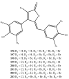
Figure 6
Figure 6
Structures of cinerols A–C (3133) and cinerol F (34).
Figure 7
Figure 7
Structures of compound 35 and 36.
Figure 8
Figure 8
Structures of frondoplysins A (37) and B (38).
Figure 9
Figure 9
Structures of nakijinol G (39) and its inactive congeners nakijinol B (40) and agelasine G (41).
Figure 10
Figure 10
Structures of sesquiterpenes 4244.
Figure 11
Figure 11
Structures of compounds 4547.
Figure 12
Figure 12
Structures of diterpenoids 4859.
Figure 13
Figure 13
Structures of strongylophorines (6066).
Figure 14
Figure 14
Structures of the diterpene-type compounds 6770.
Figure 15
Figure 15
Structure of hippolide derivatives 7177.
Figure 16
Figure 16
Structures of scalarane-type sesterterpenes: hyattellactones A and B (78 and 79), phyllofolactones F and G (80 and 81), and compounds 8283.
Figure 17
Figure 17
Structures of (7E, 12E, 20Z, 18S)-variabilin and (12E, 20Z, 18S)-8-hydroxyvariabilin (84 and 85, respectively), as well as furospongin-1 (86) and 11-O-acetylfurospongin-1 (87).
Figure 18
Figure 18
Structures of hyrtiosal (88) and stellettin G (89).
Figure 19
Figure 19
Structures of sterols 9095.
Figure 20
Figure 20
Structure of polybromodiphenyl ether derivatives 96104.
Figure 21
Figure 21
Structures of bromo-spiroalkaloids (compounds 105 and 106) and brominated lipid (compound 107).
Figure 22
Figure 22
Structure of phosphoeleganin (108) and its semisynthetic derivatives 109 and 110.
Figure 23
Figure 23
Structure of woodylide C (111).
Figure 24
Figure 24
Structures of melophlin C (112) and some active polyacetylene derivatives 113116.
Figure 25
Figure 25
Structures of 5-alkylpyrrole-2-carboxaldehyde derivatives 117123.
Figure 26
Figure 26
Structures of sulfates isolated from Penares sp. (124129).
Figure 27
Figure 27
Structures of polyacetylenic acids from marine sponges (130133).
Figure 28
Figure 28
Structures of unsaturated fatty acids 134 and 135, as well as the tricyclic spiroketal compound 136.
Figure 29
Figure 29
Structures of glycosides 137141, gallic acid (142), and its methyl ether derivative (143). Helianthamide and magnificamide are reported with disulfide connectivity, which is highlighted by the same color of bonding partners.
Figure 30
Figure 30
Structures of carteriosulfonic acids A–C (144146) and the semisynthetic derivative 147.
Figure 31
Figure 31
Structures of natural manzamine derivatives 148151.
Figure 32
Figure 32
Structure of (Z)-5-(4-hydroxybenzylidene)hydantoin (167).
Figure 33
Figure 33
Structures of palinurin (168) and of ircinin-1 and ircinin-2 (169 and 170).
Figure 34
Figure 34
Structure of meridianins 170176 and iso-meridianins 177 and 178.
Figure 35
Figure 35
Structure of nelliellosides A and B (179 and 180) and the halenaquinol sulphate (compound 181).
Figure 36
Figure 36
Structure of hymenialdisine (182), spongiacidin B (183), and leucettamine B (184).
Figure 37
Figure 37
Structures of polyandrocarpamines A and B (185 and 186), clathridine (187), and clathridimine (188).
Figure 38
Figure 38
Structures of polybrominated diphenyl ethers (compounds 189191) and alkaloids 192195.
Figure 39
Figure 39
Structure of rubrolides 196203.
Figure 40
Figure 40
Structures of spongiacidins 204210.
Figure 41
Figure 41
Structures of psammaplin A (211), compounds 212214, gracilioethers B and C (215216), and plakilactone C (217).
Figure 42
Figure 42
Structures of compounds 218222.
Figure 43
Figure 43
Structure of (−)-muqubilin A (223).
Figure 44
Figure 44
Graphical description of the main classes of MNPs and MNP-derived compounds involved in T2DM and its complications.
Figure 45
Figure 45
Numerical representation of the reported MNPs and their semisynthetic derivatives involved in T2DM and related complications. (A) Relative percentage of bioactive MNPs and MNP-derived compounds with respect to the selected targets. (B) Histogram of MNPs active on a specific target, categorized by the most representative invertebrates.

References

    1. World Health Organization. [(accessed on 17 July 2023)]. Available online: https://www.who.int/health-topics/diabetes#tab=tab_1.
    1. Shaw J.E., Sicree R.A., Zimmet P.Z. Global Estimates of the Prevalence of Diabetes for 2010 and 2030. Diabetes Res. Clin. Pract. 2010;87:4–14. doi: 10.1016/j.diabres.2009.10.007. - DOI - PubMed
    1. Nolan C.J., Damm P., Prentki M. Type 2 Diabetes across Generations: From Pathophysiology to Prevention and Management. Lancet. 2011;378:169–181. doi: 10.1016/S0140-6736(11)60614-4. - DOI - PubMed
    1. Kahn B.B., Flier J.S. Obesity and Insulin Resistance. J. Clin. Investig. 2000;106:473–481. doi: 10.1172/JCI10842. - DOI - PMC - PubMed
    1. Roberts C.K., Hevener A.L., Barnard R.J. Comprehensive Physiology. John Wiley & Sons; Hoboken, NJ, USA: 2013. Metabolic Syndrome and Insulin Resistance: Underlying Causes and Modification by Exercise Training; pp. 1–58. - PMC - PubMed

LinkOut - more resources