. 2023 Sep 16;15(9):2333.
doi: 10.3390/pharmaceutics15092333.

Autologous and Allogeneic Cytotherapies for Large Knee (Osteo)Chondral Defects: Manufacturing Process Benchmarking and Parallel Functional Qualification

Affiliations

Autologous and Allogeneic Cytotherapies for Large Knee (Osteo)Chondral Defects: Manufacturing Process Benchmarking and Parallel Functional Qualification

Virginie Philippe et al. Pharmaceutics. .

Abstract

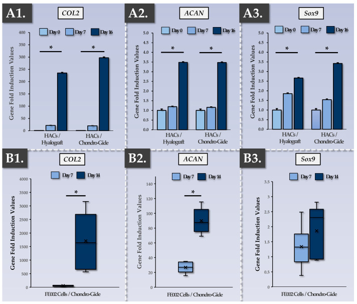
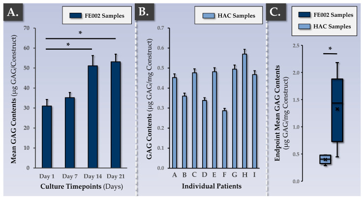
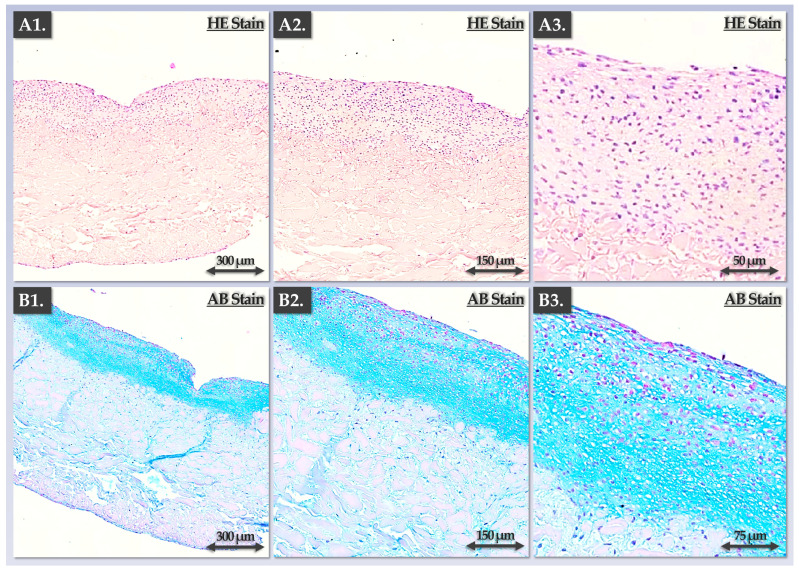
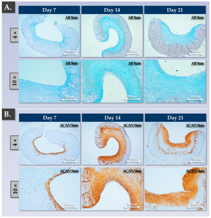
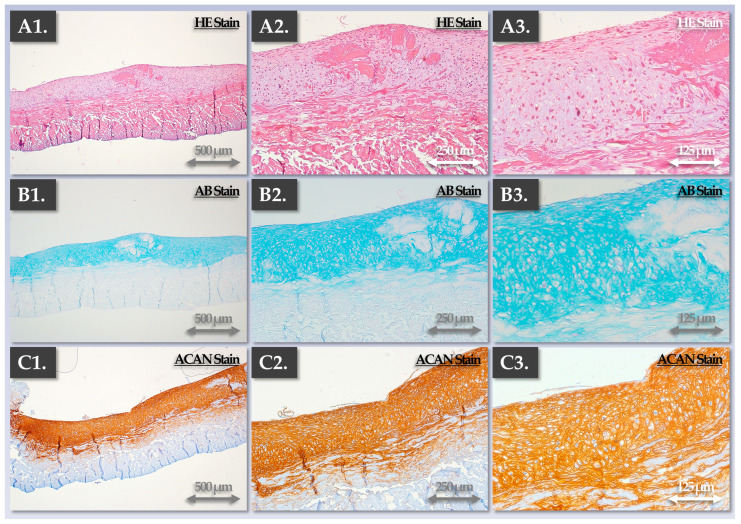
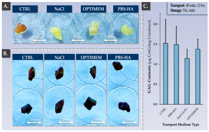
Cytotherapies are often necessary for the management of symptomatic large knee (osteo)-chondral defects. While autologous chondrocyte implantation (ACI) has been clinically used for 30 years, allogeneic cells (clinical-grade FE002 primary chondroprogenitors) have been investigated in translational settings (Swiss progenitor cell transplantation program). The aim of this study was to comparatively assess autologous and allogeneic approaches (quality, safety, functional attributes) to cell-based knee chondrotherapies developed for clinical use. Protocol benchmarking from a manufacturing process and control viewpoint enabled us to highlight the respective advantages and risks. Safety data (telomerase and soft agarose colony formation assays, high passage cell senescence) and risk analyses were reported for the allogeneic FE002 cellular active substance in preparation for an autologous to allogeneic clinical protocol transposition. Validation results on autologous bioengineered grafts (autologous chondrocyte-bearing Chondro-Gide scaffolds) confirmed significant chondrogenic induction (COL2 and ACAN upregulation, extracellular matrix synthesis) after 2 weeks of co-culture. Allogeneic grafts (bearing FE002 primary chondroprogenitors) displayed comparable endpoint quality and functionality attributes. Parameters of translational relevance (transport medium, finished product suturability) were validated for the allogeneic protocol. Notably, the process-based benchmarking of both approaches highlighted the key advantages of allogeneic FE002 cell-bearing grafts (reduced cellular variability, enhanced process standardization, rationalized logistical and clinical pathways). Overall, this study built on our robust knowledge and local experience with ACI (long-term safety and efficacy), setting an appropriate standard for further clinical investigations into allogeneic progenitor cell-based orthopedic protocols.

Keywords: FE002 primary chondroprogenitors; allogeneic cytotherapies; autologous chondrocyte implantation; cartilage defect; cell therapy; chondrogenesis; manufacturing process; standardized transplant product; tissue engineering; translational research.


Conflict of interest statement

Authors A.J., C.P. and A.L. were employed by LAM Biotechnologies SA (Epalinges, Switzerland) during the production of this work. The remaining authors declare no conflicts of interest.

Figures

Figure 1
Figure 1
Schematic presentation of the manufacturing and control processes for the autologous or allogeneic chondrogenic cellular active substances. The process describes a primary cell amplification and cryopreservation cycle, starting with master cell bank (MCB) or working cell bank (WCB) vials of HACs or FE002 primary chondroprogenitors. (A) Cellular seeding material initiation from cryopreservation with rapid thawing and culture vessel seeding. (B) In vitro cellular expansion in monolayers for cellular active substance lot manufacture. (C) Cellular bulk harvest and cellular active substance lot processing for cryopreservation. CPP, critical process parameter; HAC, human articular chondrocytes; IPC, in-process control; KPP, key process parameter; MCB, master cell bank; PPC, post-process control; WCB, working cell bank.
Figure 2
Figure 2
Schematic presentation of the manufacturing and control processes for the allogeneic finished cytotherapeutic product (i.e., Chondro-Gide scaffolds bearing FE002 primary chondroprogenitors). The process describes cellular active substance lot initiation, scaffold seeding and chondrogenic induction, and finished product conditioning for transport. (A) Cellular active substance initiation from cryopreservation with rapid thawing. It is important to note that, in the autologous protocol, an additional in vitro HAC monolayer expansion phase is carried out at this point. (B) Seeding of the cellular active substance on the Chondro-Gide scaffold and incubation of the constructs under chemical chondrogenic induction. (C) Endpoint harvest of the finished cytotherapeutic product lot and conditioning for transport to the clinical site. CPP, critical process parameter; HAC, human articular chondrocytes; IPC, in-process control; KPP, key process parameter; PPC, post-process control.
Figure 3
Figure 3
Functional characterization of the autologous and allogeneic finished products, as assessed according to their evolutive chondrogenic gene expression levels during construct incubation. (A1A3) Relative chondrogenic gene (i.e., COL2, ACAN, Sox9) fold induction values at various timepoints of autologous finished product incubation (assessed for Hyalograft and Chondro-Gide scaffolds, respectively). Both scaffolds were assessed as being functionally equivalent, and endpoint chondrogenic gene expression was highly significantly increased compared to the baseline in all groups (p-values < 0.01). (B1B3) Relative chondrogenic gene (i.e., COL2, ACAN, Sox9) fold induction values at various timepoints of allogeneic finished product incubation (assessed for Chondro-Gide scaffolds). Endpoint chondrogenic gene expression was highly significantly increased for COL2 (p-value < 0.01) and ACAN (p-value < 0.0001). Furthermore, endpoint chondrogenic gene expression was significantly higher in value for COL2 and ACAN (p-values < 0.01) compared to the respective endpoint induction levels of the same genes in the autologous finished products (i.e., the Chondro-Gide groups). Experimental replicates (n ≥ 3) and repetitions were used for the assays. Statistically significant differences are marked by an asterisk (i.e., “*”). ACAN, aggrecan; COL, collagen.
Figure 4
Figure 4
Functional characterization of the autologous and allogeneic finished products, as assessed via total GAG quantification within the constructs. (A) Increase in GAG contents over time within constructs bearing allogeneic FE002 primary chondroprogenitors. Experimental replicates (n ≥ 6) and repetitions were used for the assay. Statistically significant differences (p-values < 0.05) are marked by an asterisk (i.e., “*”). (B) Interpatient variability in terms of endpoint GAG contents within freshly harvested autologous constructs. Experimental replicates (n ≥ 3) from nine different donors were used for the assay. (C) Comparison of endpoint GAG contents between constructs bearing HACs (i.e., 16 days of induction) and constructs bearing FE002 primary chondroprogenitors (i.e., 14 days of induction). Experimental replicates (n ≥ 6) and repetitions were used for the assay. Statistically significant differences (p-value < 0.05) are marked by an asterisk (i.e., “*”). GAG, glycosaminoglycan; HAC, human articular chondrocytes.
Figure 5
Figure 5
Endpoint functional characterization of the autologous finished product (as assessed by immunohistology for constructs bearing HACs). (A1A3) Sections of a construct following HE staining. (B1B3) Sections of a construct following AB staining. The results showed the zone-specific localization of the HACs (i.e., rounded cells within lacunae) and the deposited ECM (i.e., one defined construct zone), as expected. AB, Alcian Blue; ECM, extracellular matrix; HAC, human articular chondrocytes; HE, hematoxylin and eosin.
Figure 6
Figure 6
Functional characterization (time-course) of the allogeneic finished product, as assessed via immunohistology for constructs bearing FE002 primary chondroprogenitors. (A) Construct sections at various timepoints of the incubation phase following AB staining. (B) Construct sections at various timepoints of the incubation phase following ACAN staining. Overall, the results showed that significant ECM deposition occurred between days 7 and 14 of the construct incubation phase. AB, Alcian blue; ACAN, aggrecan; ECM, extracellular matrix.
Figure 7
Figure 7
Endpoint characterization of the allogeneic finished products (as assessed via immunohistology for constructs bearing FE002 primary chondroprogenitors). In addition to significant ECM deposition in one layer of the constructs, the cells were observed to be rounded and localized in the lacunae, as expected. (A1A3) Hematoxylin and eosin staining. (B1B3) Alcian Blue staining. (C1C3) Aggrecan staining. AB, Alcian Blue; ACAN, aggrecan; ECM, extracellular matrix; HE, hematoxylin and eosin.
Figure 8
Figure 8
Validation results for allogeneic finished product transport medium and construct suturability. (A) Freshly harvested finished products before conditioning for transport/storage. Experimental replicates (n = 3) were used for the assay. (B) MTT-stained finished products after the application of the standardized transport protocol. Constructs presented in the top row were additionally submitted to the suture test before MTT staining. Experimental replicates (n = 3) were used for the assay. (C) Impact of the transport protocol on the total GAG contents of the constructs. Experimental replicates (n = 6) were used for the assay. CTRL, control; HA, hyaluronic acid; PBS, phosphate-buffered saline.

References

    1. Niemeyer P., Hanus M., Belickas J., László T., Gudas R., Fiodorovas M., Cebatorius A., Pastucha M., Hoza P., Magos K., et al. Treatment of large cartilage defects in the knee by hydrogel-based autologous chondrocyte implantation: Two-year results of a prospective, multicenter, single-arm phase III trial. Cartilage. 2022;13:19476035221085146. doi: 10.1177/19476035221085146. - DOI - PMC - PubMed
    1. Makris E.A., Gomoll A.H., Malizos K.N., Hu J.C., Athanasiou K.A. Repair and tissue engineering techniques for articular cartilage. Nat. Rev. Rheumatol. 2015;11:21–34. doi: 10.1038/nrrheum.2014.157. - DOI - PMC - PubMed
    1. Brittberg M., Gomoll A.H., Canseco J.A., Far J., Lind M., Hui J. Cartilage repair in the degenerative ageing knee. Acta Orthop. 2016;87:26–38. doi: 10.1080/17453674.2016.1265877. - DOI - PMC - PubMed
    1. Martin R., Laurent A., Applegate L.A., Philippe V. Grands défects chondraux et ostéochondraux du genou: Traitement par greffe chondrocytaire autologue. Rev. Med. Suisse. 2022;18:2384–2390. doi: 10.53738/REVMED.2022.18.808.2384. - DOI - PubMed
    1. Saris D.B., Vanlauwe J., Victor J., Almqvist K.F., Verdonk R., Bellemans J., Luyten F.P., TIG/ACT/01/2000&EXT Study Group Treatment of symptomatic cartilage defects of the knee: Characterized chondrocyte implantation results in better clinical outcome at 36 months in a randomized trial compared to microfracture. Am. J. Sports Med. 2009;37:10S–19S. doi: 10.1177/0363546509350694. - DOI - PubMed

LinkOut - more resources