Review
. 2023 Sep 21;15(18):3849.
doi: 10.3390/polym15183849.

Advancements in Chitosan-Based Nanoparticles for Pulmonary Drug Delivery

Affiliations
Review

Advancements in Chitosan-Based Nanoparticles for Pulmonary Drug Delivery

Thiago Medeiros Zacaron et al. Polymers (Basel). .

Abstract

The evolution of respiratory diseases represents a considerable public health challenge, as they are among the leading causes of death worldwide. In this sense, in addition to the high prevalence of diseases such as asthma, chronic obstructive pulmonary disease, pneumonia, cystic fibrosis, and lung cancer, emerging respiratory diseases, particularly those caused by members of the coronavirus family, have contributed to a significant number of deaths on a global scale over the last two decades. Therefore, several studies have been conducted to optimize the efficacy of treatments against these diseases, focusing on pulmonary drug delivery using nanomedicine. Thus, the development of nanocarriers has emerged as a promising alternative to overcome the limitations of conventional therapy, by increasing drug bioavailability at the target site and reducing unwanted side effects. In this context, nanoparticles composed of chitosan (CS) show advantages over other nanocarriers because chitosan possesses intrinsic biological properties, such as anti-inflammatory, antimicrobial, and mucoadhesive capacity. Moreover, CS nanoparticles have the potential to enhance drug stability, prolong the duration of action, improve drug targeting, control drug release, optimize dissolution of poorly soluble drugs, and increase cell membrane permeability of hydrophobic drugs. These properties could optimize the performance of the drug after its pulmonary administration. Therefore, this review aims to discuss the potential of chitosan nanoparticles for pulmonary drug delivery, highlighting how their biological properties can improve the treatment of pulmonary diseases, including their synergistic action with the encapsulated drug.

Keywords: chitosan; chitosan derivatives; lung delivery; lung diseases; nanoparticles; pulmonary drug delivery.


Conflict of interest statement

The authors declare no conflict of interest.

Figures

Figure 1
Figure 1
Alkaline reaction for the deacetylation of chitin with the formation of CS. Generally, chitin undergoes a treatment process in which it is exposed to a concentrated NaOH solution at elevated temperatures for an extended period of time, producing chitosan as an insoluble residue.
Figure 2
Figure 2
Schematic representation of the chitosan/TPP interaction in the ionic gelation process. The interaction occurs between the protonated amino groups of chitosan and the negative charges of TPP after its ionization in an aqueous medium.
Figure 3
Figure 3
Examples of some CS-based nanoparticles, which can be essentially formed by CS or prepared from CS-coated phospholipidic, lipidic or polymeric nanoparticles. (A): CS nanosphere, with drug dispersed in polymer matrix; (B): CS-coated liposome. In this case, hydrophilic drugs can be encapsulated in the aqueous compartment, hydrophobic drugs in lipid bilayers and amphiphilic drugs between these two compartments.; (C): CS-coated solid lipid nanoparticle with the drug encapsulated in the oil core; and (D): CS-coated polymeric nanocapsule, where the drug can be encapsulated within the oil core or adsorbed to the polymeric wall.
Figure 4
Figure 4
Some important advantages of CS nanoparticles (A) in pulmonary drug delivery. Inhaled CS nanoparticles are able to improve antiviral (B) and antibacterial (C) activities, facilitate drug penetration through the mucus layer (D), contribute to anti-inflammatory activity (E), and increase the interaction/internalization in specific cells such as macrophages (F) and tumor cells (G).
Figure 5
Figure 5
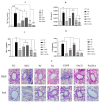
Effect of FACHA on serum levels of IgE (A), IL5 (B), TNF-α (C) and IL13 (D) quantified using ELISA after pulmonary administration in an OVA-induced mouse model of asthma. These cytokines play distinct but related roles in the pathogenesis of asthma, contributing to the inflammation, bronchial hyperreactivity, and excessive mucus production that characterize the disease. (A) shows that, compared to FACHA treatment, FA demonstrated a more limited ability to reduce serum IgE levels. Also, FACHA treatment showed remarkable efficacy (p ≤ 0.001) in reducing IL5 (B), TNFα (C), and IL13 ((D) levels. This provides compelling evidence for the ability of the formulation to alleviate the asthmatic condition in OVA-sensitized mice. (E) shows histopathologic sections of lungs from treated mice. As can be seen, the FACHA-treated group showed normal morphological features, suggesting its protective effect against excessive mucus secretion and inflammatory cell infiltration in lung tissue. These findings support the enhanced efficacy of FACHA compared to pure FA, which is attributed to the mucoadhesive nanocarrier properties that enhance drug retention and facilitate transport across the pulmonary barrier. NC: normal control (0.9 N saline); NE: negative control (OVA sensitization); PC: positive control (OVA sensitization followed by budesonide treatment (9.50 mg/m3); FA: ferulic acid; CSNP: unloaded CS nanoparticles; FACS: ferulic acid-loaded CS nanoparticle; FACHA: hyaluronic acid functionalized ferulic acid-loaded CS nanoparticle.; H&E: hematoxylin and eosin; PAS: periodic acid Schiff stain; Statistical significance—*** (p ≤ 0.001), ** (p ≤ 0.01), * (p ≤ 0.05), ns (p greater than 0.05) [18]. Reproduced with permission from Dhayanandamoorthy et al., Int. J. Pharm.; published by Elsevier, 2020.
Figure 6
Figure 6
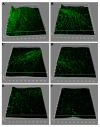
Confocal laser scanning microscopic images of P. aeruginosa biofilm after different treatments: untreated (A), CIPR (B), CIPR + AgLase (C), CIPR-CH-NPs (D), CIPR-CH-NPs + AgLase (E) and AgLase-CIPR-CH-NPs (F). AgLase-CIPR-CH-NPs clearly showed the most potent antibiofilm activity. CIPR: ciprofloxacin; CH: chitosan; AgLase: alginate lyase; NPs: nanoparticles; CIPR-CH-NPs: ciprofloxacin-loaded chitosan nanoparticles; AgLase-CIPR-CH-NPs: alginate lyase functionalized chitosan nanoparticles of ciprofloxacin [20]. Reproduced with permission from Patel et al., Int. J. Pharm.; published by Elsevier, 2019.
Figure 7
Figure 7
Photos of lungs collected from mice after 15 days of different treatments (A). Number of lung metastatic foci on the lung surface after treatments (B). In contrast to the other groups, mice treated with CS/aPD-L1 showed a marked decrease in the quantity of lesions. This strongly suggests that the inhalation of CS/aPD-L1 effectively inhibits lung metastasis. PBS: phosphate-buffered saline. CS: chitosan; aPD-L1: anti-programmed cell death protein ligand 1; i.v.: intravenous; * p < 0.05 [128]. Reproduced with permission from Jin et al., Adv. Mater.; published by John Wiley and Sons, 2021.
Figure 8
Figure 8
Percentage of the in vivo pulmonary deposition of (a) isoniazid, (b) pyrazinamide, (c) and rifampicin after intratracheal administration of the DPI formulation compared to oral administration of each drug. (values expressed as mean ± standard deviation, n = 3). The DPI formulation maintains elevated drug levels in the lungs for a longer period of time compared to standard oral dosing [146]. Reproduced with permission from Chogale et al., Drug Deliv. Transl. Res.; published by Springer Nature, 2021.
Figure 9
Figure 9
Images of dissected mice, highlighting the alveoli (AF). (A) Control. (B) Oleic acid induced model. (C) Treatment with free capsules. (D) Treatment with nanoparticles containing extract of chamomile flowers + CUR. (E) Treatment with nanoparticles containing SIL. + CUR. Imagens of individual lungs (FJ). The arrows indicate significant functional changes. (F) Control. (G) Oleic acid-induced model. (H) Treatment with free capsules. (I) Treatment with nanoparticles containing extract of chamomile flowers + CUR. (J) Treatment with nanoparticles containing SIL. + CUR. Histopathological analysis. (F) Control. (G) Oleic acid-induced model. (H) Treatment with free capsules. (I) Treatment with nanoparticles containing extract of chamomile flowers + CUR. (J) Treatment with nanoparticles containing SIL. + CUR. (K) Control. (L) Oleic acid model. (M) animal treated by free capsules. (N) Animal treated by Encap. Cham. + CUR. (Q) Animal treated by Encap. SIL. + CUR. SIL.: silymarin CUR.: curcumin [133]. The co-encapsulation of SIL. + CUR. completely changed the histologic profile and improved the tissue histoarchitecture. Reproduced with permission from Hanafy et al., Int. J. Biol. Macromol.; published by Elsevier, 2022.

Similar articles

Cited by

References

    1. McGonagle D., Sharif K., O’Regan A., Bridgewood C. The Role of Cytokines including Interleukin-6 in COVID-19 induced Pneumonia and Macrophage Activation Syndrome-Like Disease. Autoimmun. Rev. 2020;19:102537. doi: 10.1016/j.autrev.2020.102537. - DOI - PMC - PubMed
    1. Gibson P.G., Qin L., Puah S.H. COVID-19 acute respiratory distress syndrome (ARDS): Clinical features and differences from typical pre-COVID-19 ARDS. Med. J. Aust. 2020;213:54–56. doi: 10.5694/mja2.50674. - DOI - PMC - PubMed
    1. Aveyard P., Gao M., Lindson N., Hartmann-Boyce J., Watkinson P., Young D., Coupland C.A.C., Tan P.S., Clift A.K., Harrison D., et al. Association between pre-existing respiratory disease and its treatment, and severe COVID-19: A population cohort study. Lancet Respir. Med. 2021;9:909–923. doi: 10.1016/S2213-2600(21)00095-3. - DOI - PMC - PubMed
    1. Fei Q., Bentley I., Ghadiali S.N., Englert J.A. Pulmonary drug delivery for acute respiratory distress syndrome. Pulm. Pharmacol. Ther. 2023;79:102196. doi: 10.1016/j.pupt.2023.102196. - DOI - PMC - PubMed
    1. Newman S.P. Drug delivery to the lungs: Challenges and opportunities. Ther. Deliv. 2017;8:647–661. doi: 10.4155/tde-2017-0037. - DOI - PubMed

LinkOut - more resources