

Surface Acoustic Wave Immunosensor for Detection of Botulinum Neurotoxin
- PMID: 37765744
- PMCID: PMC10534944
- DOI: 10.3390/s23187688
Surface Acoustic Wave Immunosensor for Detection of Botulinum Neurotoxin
Abstract
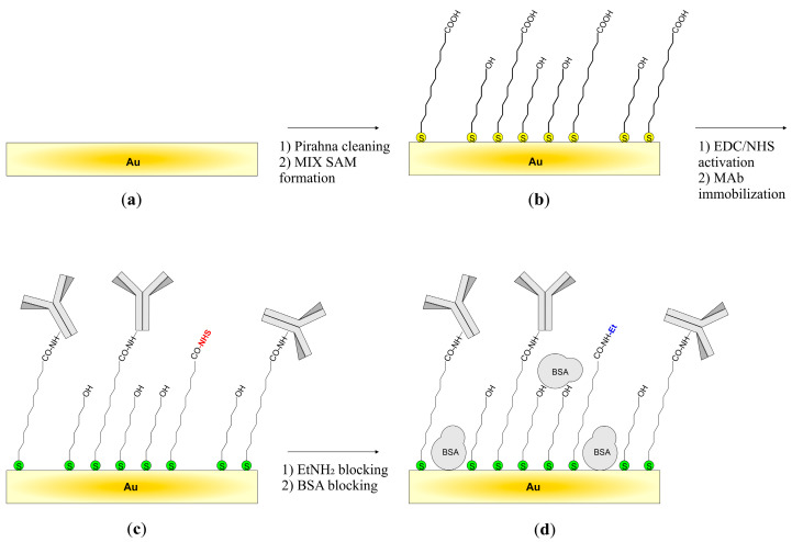
A Love-type acoustic wave sensor (AT-cut quartz substrate, SiO2 guiding layer) with a center frequency of approximately 120 MHz was used to detect a simulant of pathogenic botulinum neurotoxin type A-recombinant of BoNT-A light chain-in liquid samples. The sensor was prepared by immobilizing monoclonal antibodies specific for botulinum neurotoxin via a thiol monolayer deposited on a gold substrate. Studies have shown that the sensor enables selective analyte detection within a few minutes. In addition, the sensor can be used several times (regeneration of the sensor is possible using a low pH buffer). Nevertheless, the detectability of the analyte is relatively low compared to other analytical techniques that can be used for rapid detection of botulinum neurotoxin. The obtained results confirm the operation of the proposed sensor and give hope for further development of this label-free technique for detecting botulinum neurotoxin.
Keywords: SAW; biological aerosol; biological warfare agents (BWA); botulinum neurotoxin; label-free detection; on-site detection of BWA.
Conflict of interest statement
The authors declare no conflict of interest. The funders had no role in the design of the study; in the collection, analyses, or interpretation of data; in the writing of the manuscript, or in the decision to publish the results.
Figures


Similar articles
-
Highly Sensitive Love Mode Acoustic Wave Platform with SiO2 Wave-Guiding Layer and Gold Nanoparticles for Detection of Carcinoembryonic Antigens.Biosensors (Basel). 2022 Jul 18;12(7):536. doi: 10.3390/bios12070536. Biosensors (Basel). 2022. PMID: 35884339 Free PMC article.
-
Fabrication of a Novel Highly Sensitive and Selective Immunosensor for Botulinum Neurotoxin Serotype A Based on an Effective Platform of Electrosynthesized Gold Nanodendrites/Chitosan Nanoparticles.Sensors (Basel). 2017 May 9;17(5):1074. doi: 10.3390/s17051074. Sensors (Basel). 2017. PMID: 28486408 Free PMC article.
-
Surface acoustic wave immunosensor for real-time detection of hepatitis B surface antibodies in whole blood samples.Biosens Bioelectron. 2009 Jun 15;24(10):3120-5. doi: 10.1016/j.bios.2009.04.009. Epub 2009 Apr 17. Biosens Bioelectron. 2009. PMID: 19423329
-
Botulinum neurotoxin: where are we with detection technologies?Crit Rev Microbiol. 2013 Feb;39(1):43-56. doi: 10.3109/1040841X.2012.691457. Epub 2012 Jun 8. Crit Rev Microbiol. 2013. PMID: 22676403 Review.
-
A High Fundamental Frequency (HFF)-based QCM Immunosensor for Tuberculosis Detection.Curr Top Med Chem. 2017;17(14):1623-1630. doi: 10.2174/1568026617666161104105210. Curr Top Med Chem. 2017. PMID: 27823567 Review.
Cited by
-
Wearable Temperature Sensor Enhanced Volatilomics Technique for Swift and Convenient Detection of Latrogenic Botulism.Adv Sci (Weinh). 2025 Feb;12(6):e2411738. doi: 10.1002/advs.202411738. Epub 2024 Dec 16. Adv Sci (Weinh). 2025. PMID: 39679864 Free PMC article.
References
-
- Gupta R. Handbook of Toxicology of Chemical Warfare Agents. Elsevier; Amsterdam, The Netherlands: 2020.
-
- CDC|Bioterrorism Agents/Diseases (by Category) [(accessed on 8 August 2023)]; Available online: https://emergency.cdc.gov/agent/agentlist-category.asp.
MeSH terms
Substances
Grants and funding
LinkOut - more resources
Full Text Sources
