. 2023 Aug 23;11(9):1404.
doi: 10.3390/vaccines11091404.

Oral Immunization with rVSV Bivalent Vaccine Elicits Protective Immune Responses, Including ADCC, against Both SARS-CoV-2 and Influenza A Viruses

Affiliations

Oral Immunization with rVSV Bivalent Vaccine Elicits Protective Immune Responses, Including ADCC, against Both SARS-CoV-2 and Influenza A Viruses

Maggie Jing Ouyang et al. Vaccines (Basel). .

Abstract

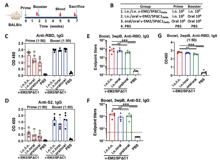
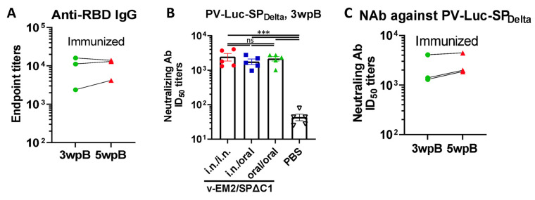
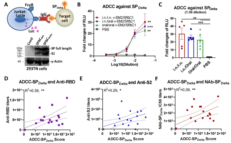
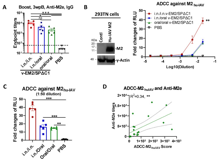
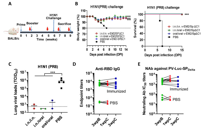
COVID-19 and influenza both cause enormous disease burdens, and vaccines are the primary measures for their control. Since these viral diseases are transmitted through the mucosal surface of the respiratory tract, developing an effective and convenient mucosal vaccine should be a high priority. We previously reported a recombinant vesicular stomatitis virus (rVSV)-based bivalent vaccine (v-EM2/SPΔC1Delta) that protects animals from both SARS-CoV-2 and influenza viruses via intramuscular and intranasal immunization. Here, we further investigated the immune response induced by oral immunization with this vaccine and its protective efficacy in mice. The results demonstrated that the oral delivery, like the intranasal route, elicited strong and protective systemic immune responses against SARS-CoV-2 and influenza A virus. This included high levels of neutralizing antibodies (NAbs) against SARS-CoV-2, as well as strong anti-SARS-CoV-2 spike protein (SP) antibody-dependent cellular cytotoxicity (ADCC) and anti-influenza M2 ADCC responses in mice sera. Furthermore, it provided efficient protection against challenge with influenza H1N1 virus in a mouse model, with a 100% survival rate and a significantly low lung viral load of influenza virus. All these findings provide substantial evidence for the effectiveness of oral immunization with the rVSV bivalent vaccine.

Keywords: ADCC activity; COVID-19; SARS-CoV-2; VSV vector; bivalent vaccine; influenza virus; neutralizing antibody; oral immunization; vesicular stomatitis virus (VSV).


Conflict of interest statement

The authors declare no conflict of interest.

Figures

Figure 1
Figure 1
Oral immunization with vaccine candidate v-EM2/SPΔC1Delta elicited robust anti-SARS-CoV-2 RBD and anti-S2 immune response in mice. (A) Schematic of the immunization of bivalent rVSV vaccine candidate v-EM2/SPΔC1Delta in mice. BALB/c mice (n = 5–8/group) were immunized with v-EM2/SPΔC1Delta via oral cavity or intranasal routes at weeks 0 and 2, as indicated. The blood from mice in each group was collected on weeks 2 and 5, the mice were sacrificed at week 7 (i.e., 5 weeks post-booster (5 wpB)), and the blood was collected. (B) The immunization groups are shown with the delivery route and vaccine dose. (CF) Sera after prime (week 2) and booster (week 5, i.e., 3 weeks post-booster (3 wpB)) immunization were measured for anti-SARS-CoV-2 RBD IgG (C,E), IgA (G) levels, and anti-S2 IgG (D,F) in OD450 or endpoint titers. Data represent mean ± SEM. Statistical significance was determined using a one-way ANOVA test and Tukey’s test. ***, p < 0.001, ns: no significance.
Figure 2
Figure 2
The duration and neutralization of antibodies against SARS-CoV-2 SP induced by oral immunization with v-EM2/SPΔC1Delta in mice. (A) The duration of anti-SARS-CoV-2 RBD (endpoint titers) in the i.n. immunized mice sera after booster immunization (3 wpB and 5 wpB) were measured. (B) The neutralization titers (50% inhibitory dose, ID50) in immunized mice sera (3 wpB) against pseudovirus PV-Luc-SPDelta infection were determined. The serially diluted mouse sera were incubated with PV-Luc-SPDelta (≈104 RLU), and then the mixtures (PV + Sera) were used to inoculate A549ACE2 cells. The infection of PV was determined by luciferase assay at 48~66 h post-infection. The percentage of infection was calculated compared with the no serum control. The 50% inhibition dose (ID50) neutralizing Ab titers were calculated by using sigmoid 4PL interpolation with GraphPad Prism 9.0, as described in the Materials and Methods. (C) The duration of neutralizing Ab ID50 titers in the i.n. immunized mice sera collected at 3 wpB and 5 wpB. Data represent mean ± SEM. Statistical significance was determined using a one-way ANOVA test and Tukey’s test. ***, p < 0.001. ns: no significance. wpB, week post-boost.
Figure 3
Figure 3
Oral immunization with v-EM2/SPΔC1Delta induced the ADCC activities against SARS-CoV-2 SPDelta in mice. (A) The schematic diagram of Jurkat–Lucia-cell-based ADCC activity reporter assay as described in the Materials and Methods. The SPDelta-expressing cells were obtained from transfection and incubated with serially diluted mouse serum (containing SP-binding IgG). Jurkat–Lucia reporter cells expressing Fcγ receptor (FcγR) were added to the mixture of SPDelta-expressing cells and serum. The engagement of FcγR, IgG, and SP triggered the activation of Jurkat effector cells and the luciferase (Luc) expression. The ADCC activity was determined by measuring the secreted Luc in the cell culture supernatant (upper). The antigen SARS-CoV-2 spike protein (SPΔCWH, SPΔCDelta) expression in target 293TN cells was determined by Western blotting (lower panel) at 24 h post-transfection using the anti-SARS-CoV-2 SP-NTD (Elabscience, Cat# E-AB-V1030). (B) The ADCC activities against SARS-CoV-2 SPDelta in the immunized mice sera (3 wpB) were determined as described in (A). The ADCC induction (fold change) of sera from each group was calculated against the no-serum control. (C) The ADCC against SPDelta in individual mouse serum at 1:30 dilution. Data represent mean ± SEM. Statistical significance (B,C) was determined using an ordinary one-way (C) or two-way (B) ANOVA test and Tukey’s test. (DF) The ADCC activity against SPDelta was positively correlated with the titers of anti-SP antibodies (RBD- or S2-binding) (D,E) or NAbs (F) of mice in all groups. The ADCC score was calculated as (fold-change × dilution factor). The ADCC score geomean of the first two dilutions was used. The correlation analysis was performed by Prism. The correlation coefficient (R2) and two-tailed p-value were calculated via the Pearson method by Prism. *, p < 0.05; **, p < 0.01; ***, p < 0.001.
Figure 4
Figure 4
Oral immunization with vaccine candidate v-EM2/SPΔC1Delta effectively triggered a humoral immune response against the influenza virus in mice. (A) The anti-influenza virus matrix-2 (M2) ectodomain (M2e) IgG levels in the sera of the immunized mice at 3 weeks post-booster (3 wpB) as described in Figure 1A were measured by ELISA. (B) The ADCC activities (fold change of RLU) against human influenza virus M2 (M2hu-IAV) in the immunized mice sera (3 wpB) were determined as described in Figure 3A, except using 293TN-M2 target cells. The human influenza A virus (IAV) M2 protein expression in target cells was determined by Western blotting using the sera of mice immunized with v-EM2/ SPΔC1Delta (containing anti-M2 antibodies) (left panel). The ADCC induction (fold change) of serum from each mouse was calculated against the no-serum control (right panel). (C) The ADCC against M2hu-IAV in individual mouse serum at 1:50 dilution. Data represent mean ± SEM. Statistical significance was determined using one-way (A,C) or two-way (B) ANOVA test and Tukey’s test. (D) The correlation of ADCC activity against M2hu-IAV and the titers of anti-M2e of mice in all groups. The ADCC score was calculated as described in Figure 3D. The correlation analysis was performed by Prism. The correlation coefficient (R2) and two-tailed p-value were calculated via the Pearson method. *, p < 0.05; **, p < 0.01; ***, p < 0.001. ns: no significance. wpB, week post-boost.
Figure 5
Figure 5
Oral immunization with v-EM2/SPΔC1Delta protected mice from the lethal challenge of H1N1 influenza virus. (A) Schematic of the immunization of bivalent rVSV vaccine v-EM2/SPΔC1Delta and influenza challenge in mice. BALB/c mice (5/group) were immunized with v-EM2/SPΔC1Delta via oral cavity or intranasal routes. At week 6, i.e., 4 weeks post-boost, mice of each group were intranasally infected with the H1N1 influenza virus PR8 (3 × 10³ TCID50). Then, the percentages of original body weight ((B), left panel) and survival rates ((B), right panel) of the mice were monitored daily for 2 weeks. (C) Viral loads in the lung tissues of immunized mice (2/group) and PBS group (n = 5) at 7 days after H1N1 challenge (1 wpC) were determined with MDCK cells, as described in the Materials and Methods. (D,E) The anti-RBD IgG endpoint titers (D) and NAb titers against PV-Luc-SPDelta in mice sera before (3 wpB) and after challenge (1 wpC and 3 wpC). The titers from the same mouse were linked with a line. Data shown are mean ± SEM. Statistical significance was determined using unpaired Student’s t-test (C) or one-way ANOVA test and Tukey’s test (A,B) ***, p < 0.001. wpB, week post-boost. wpC, week post-challenge. TCID50, 50% tissue culture infectious dose.

Similar articles

Cited by

References

    1. Andrews N., Stowe J., Kirsebom F., Toffa S., Rickeard T., Gallagher E., Gower C., Kall M., Groves N., O’Connell A.-M., et al. COVID-19 Vaccine Effectiveness against the Omicron (B.1.1.529) Variant. N. Engl. J. Med. 2022;386:1532–1546. doi: 10.1056/NEJMoa2119451. - DOI - PMC - PubMed
    1. Buchan S.A., Chung H., Brown K.A., Austin P.C., Fell D.B., Gubbay J.B., Nasreen S., Schwartz K.L., Sundaram M.E., Tadrous M., et al. Estimated Effectiveness of COVID-19 Vaccines Against Omicron or Delta Symptomatic Infection and Severe Outcomes. JAMA Netw. Open. 2022;5:e2232760. doi: 10.1001/jamanetworkopen.2022.32760. - DOI - PMC - PubMed
    1. World Health Organization WHO Coronavirus (COVID-19) Dashboard. [(accessed on 15 June 2023)];2023 Available online: https://covid19.who.int/table.
    1. Tang J., Zeng C., Cox T.M., Li C., Son Y.M., Cheon I.S., Wu Y., Behl S., Taylor J.J., Chakaraborty R., et al. Respiratory mucosal immunity against SARS-CoV-2 after mRNA vaccination. Sci. Immunol. 2022;7:eadd4853. doi: 10.1126/sciimmunol.add4853. - DOI - PMC - PubMed
    1. Azzi L., Dalla Gasperina D., Veronesi G., Shallak M., Maurino V., Baj A., Gianfagna F., Cavallo P., Dentali F., Tettamanti L., et al. Mucosal immune response after the booster dose of the BNT162b2 COVID-19 vaccine. eBioMedicine. 2023;88:104435. doi: 10.1016/j.ebiom.2022.104435. - DOI - PMC - PubMed

LinkOut - more resources