

Long COVID Complicated by Fatal Cytomegalovirus and Aspergillus Infection of the Lungs: An Autopsy Case Report
- PMID: 37766216
- PMCID: PMC10535245
- DOI: 10.3390/v15091810
Long COVID Complicated by Fatal Cytomegalovirus and Aspergillus Infection of the Lungs: An Autopsy Case Report
Abstract
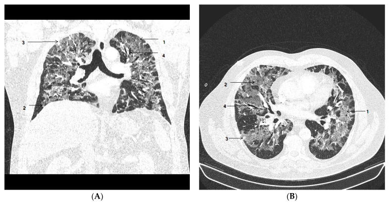
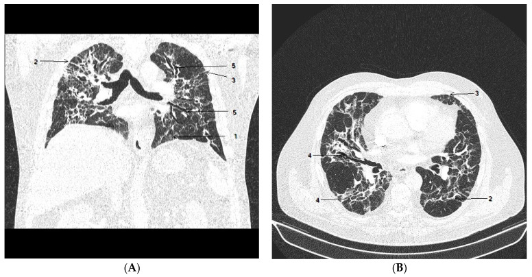
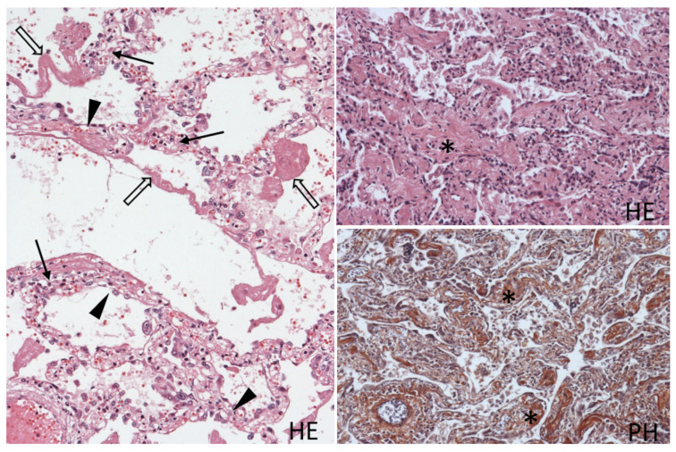
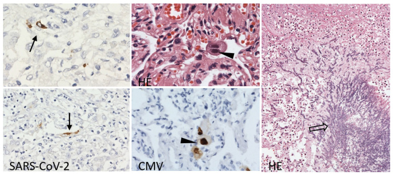
After the acute phase of COVID-19, some patients develop long COVID. This term is used for a variety of conditions with a complex, yet not fully elucidated etiology, likely including the prolonged persistence of the virus in the organism and progression to lung fibrosis. We present a unique autopsy case of a patient with severe COVID-19 with prolonged viral persistence who developed interstitial lung fibrosis complicated by a fatal combination of cytomegalovirus and Aspergillus infection. SARS-CoV-2 virus was detected at autopsy in the lungs more than two months after the acute infection, although tests from the nasopharynx were negative. Immune dysregulation after COVID-19 and the administration of corticoid therapy created favorable conditions for the cytomegalovirus and Aspergillus infection that were uncovered at autopsy. These pathogens may represent a risk for opportunistic infections, complicating not only the acute coronavirus infection but also long COVID, as was documented in the presented case.
Keywords: Aspergillus; COVID-19; corticoid therapy; cytomegalovirus; immune dysregulation; long COVID.
Conflict of interest statement
The authors declare no conflict of interest.
Figures
Similar articles
-
Autopsy analysis reveals increased macrophage infiltration and cell apoptosis in COVID-19 patients with severe pulmonary fibrosis.Pathol Res Pract. 2023 Dec;252:154920. doi: 10.1016/j.prp.2023.154920. Epub 2023 Nov 4. Pathol Res Pract. 2023. PMID: 37948998
-
Pulmonary pathology of severe acute respiratory syndrome in Toronto.Mod Pathol. 2005 Jan;18(1):1-10. doi: 10.1038/modpathol.3800247. Mod Pathol. 2005. PMID: 15272286 Free PMC article.
-
[Lung pathology in post-covid syndrome].Arkh Patol. 2023;85(5):52-59. doi: 10.17116/patol20238505152. Arkh Patol. 2023. PMID: 37814851 Russian.
-
Molecular Pathogenesis of Fibrosis, Thrombosis and Surfactant Dysfunction in the Lungs of Severe COVID-19 Patients.Biomolecules. 2022 Dec 10;12(12):1845. doi: 10.3390/biom12121845. Biomolecules. 2022. PMID: 36551272 Free PMC article. Review.
-
Pathogenesis-directed therapy of 2019 novel coronavirus disease.J Med Virol. 2021 Mar;93(3):1320-1342. doi: 10.1002/jmv.26610. Epub 2020 Nov 10. J Med Virol. 2021. PMID: 33073355 Review.
Cited by
-
Tropheryma whipplei infection in the lung of a patient with long COVID: a case report.BMC Infect Dis. 2024 Mar 6;24(1):292. doi: 10.1186/s12879-024-09183-6. BMC Infect Dis. 2024. PMID: 38448808 Free PMC article.
-
Clinical and molecular landscape of prolonged SARS-CoV-2 infection with resistance to remdesivir in immunocompromised patients.PNAS Nexus. 2025 Mar 18;4(4):pgaf085. doi: 10.1093/pnasnexus/pgaf085. eCollection 2025 Apr. PNAS Nexus. 2025. PMID: 40160532 Free PMC article.
References
-
- Krivosíkova L., Horak S., Mikus-Kuracinova K., Janega P., Palkovic M., Babal P. Contribution of immunohistochemistry to histomorphological diagnostics of COVID-19 pneumonia. Newslab. 2021;12:12–16.
-
- Phetsouphanh C., Darley D.R., Wilson D.B., Howe A., Munier C.M.L., Patel S.K., Juno J.A., Burrell L.M., Kent S.J., Dore G.J., et al. Immunological dysfunction persists for 8 months following initial mild-to-moderate SARS-CoV-2 infection. Nat. Immunol. 2022;23:210–216. doi: 10.1038/s41590-021-01113-x. - DOI - PubMed
-
- Alanio C., Verma A., Mathew D., Gouma S., Liang G., Dunn T., Oldridge D.A., Weaver J., Kuri-Cervantes L., Pampena M.B., et al. Cytomegalovirus latent infection is associated with an increased risk of COVID-19-related hospitalization. J. Infect. Dis. 2022;226:463–473. doi: 10.1093/infdis/jiac020. - DOI - PMC - PubMed
Publication types
MeSH terms
LinkOut - more resources
Full Text Sources
Medical
Miscellaneous
