. 2023 Aug 25;15(9):1813.
doi: 10.3390/v15091813.

Targeting FOXP3 Tumor-Intrinsic Effects Using Adenoviral Vectors in Experimental Breast Cancer

Affiliations

Targeting FOXP3 Tumor-Intrinsic Effects Using Adenoviral Vectors in Experimental Breast Cancer

Alejandro J Nicola Candia et al. Viruses. .

Abstract

The regulatory T cell master transcription factor, Forkhead box P3 (Foxp3), has been detected in cancer cells; however, its role in breast tumor pathogenesis remains controversial. Here we assessed Foxp3 tumor intrinsic effects in experimental breast cancer using a Foxp3 binder peptide (P60) that impairs Foxp3 nuclear translocation. Cisplatin upregulated Foxp3 expression in HER2+ and triple-negative breast cancer (TNBC) cells. Foxp3 inhibition with P60 enhanced chemosensitivity and reduced cell survival and migration in human and murine breast tumor cells. We also developed an adenoviral vector encoding P60 (Ad.P60) that efficiently transduced breast tumor cells, reduced cell viability and migration, and improved the cytotoxic response to cisplatin. Conditioned medium from transduced breast tumor cells contained lower levels of IL-10 and improved the activation of splenic lymphocytes. Intratumoral administration of Ad.P60 in breast-tumor-bearing mice significantly reduced tumor infiltration of Tregs, delayed tumor growth, and inhibited the development of spontaneous lung metastases. Our results suggest that Foxp3 exerts protumoral intrinsic effects in breast cancer cells and that gene-therapy-mediated blockade of Foxp3 could constitute a therapeutic strategy to improve the response of these tumors to standard treatment.

Keywords: Foxp3; breast cancer; cell penetrating peptide; chemosensitivity; gene therapy.


Conflict of interest statement

The authors declare no conflict of interest.

Figures

Figure 1
Figure 1
Regulation of Foxp3 expression. Foxp3 expression was assessed by flow cytometry in LM3 cells incubated for 24 h in the presence of 10 ng/mL of recombinant TGF-β (A), 50 μM of the mTOR pathway inhibitor rapamycin (RAPA) (B), or 50 μM of the COX-2 inhibitor indomethacin (INDO) (C). Representative histograms are shown. The histograms of cells incubated with isotype controls are shown in gray. * p < 0.05 vs control (Student’s t-test). (D) TNBC subtype EO771 breast cancer cells and (E) HER2+ LM3 breast cancer cells were incubated with 2 or 20 μM cisplatin for 48 h and then Foxp3 expression was evaluated by flow cytometry. Representative histograms are shown. * p < 0.05, ANOVA.
Figure 2
Figure 2
Foxp3 tumor intrinsic effects in breast cancer cells: chemoresistance. (A) TNBC EO771 and (C) HER2+ LM3 cells were incubated with peptide P60 or P301 at concentrations of 12.5, 25, or 50 μM in the presence or absence of 5 μM cisplatin for 72 h. Cell viability was determined by MTT assay. Viability values were expressed as a percentage of control without any treatment. * p < 0.05 vs. respective control, ˆ p < 0.05 vs. peptide concentrations, 2-way ANOVA. (B) EO771 and (D) LM3 cells were incubated with P301 or P60 (50 μM) in combination with 5 μM cisplatin and viability (by MTT assay) and cell proliferation (by BrdU incorporation) were evaluated 72 h later. * p < 0.05 vs. P301; ^ p < 0.05 vs. respective control without cisplatin, 2-way ANOVA. (E) The activity of caspase 3 was measured by fluorescence intensity in LM3 cells incubated for 48 h with P301 or P60 (50 μM) and cisplatin (5 μM). (F) LM3 cells were incubated for 48 h with P301 or P60 (50 μM) and cisplatin (5 μM), and the percentage of dead cells was determined by propidium iodide incorporation measured by flow cytometry. Representative histograms are shown and depict the percentage of dead cells. * p < 0.05 vs. P301; ^ p < 0.05 vs. respective control without cisplatin, 2-way ANOVA. (G) The clonogenic capacity of LM3 cells was assessed 72 h after the incubation with P301 or P60 (50 μM) in the presence or absence of cisplatin (2 μM). * p < 0.05 vs. P301; ^ p < 0.05 vs. respective control without cisplatin, 2-way ANOVA.
Figure 3
Figure 3
Foxp3 tumor intrinsic effects in breast cancer cells: invasion. (A,B) LM3 breast cancer cells were incubated for 24 h with P301 or P60 (50 μM) (A) or with the conditioned medium of cells previously incubated for 48 h with these peptides (B). The wound assay was performed and the migratory capacity of the cells evaluated. Representative microphotographs of the wound at different time points are shown. * p < 0.05, non-linear regression analysis. (C,D) The MMP-2 and MMP-9 activity was assessed by zymography in incubation media of EO771 cells (C) and LM3 cells (D) treated with P301 or P60 (50 μM) for 48 h. The gels were stained with Coomassie blue, and the bands were analyzed by densitometry with the ImageJ software in triplicate. Zymographic activity was expressed as a percentage relative to a standard internal sample saturating at a density of 50%. Representative bands for each group are shown.
Figure 4
Figure 4
Foxp3 expression and response to chemotherapy in human breast cancer cells. (A,B) Expression of Foxp3 in MDA-MB-231 (A) and MDA-MB-468 cells (B) after incubation with 40 μM or 4 μM cisplatin, respectively, was assessed by indirect immunofluorescence. Representative microphotographs show Foxp3 expression (green). Nuclei were counterstained with DAPI (blue). The percentage of cells with high intensity nuclear Foxp3 and intensity of fluorescence, expressed as corrected total cell fluorescence (CTCF), was assessed using ImageJ software. * p < 0.05 (Student’s t test). (C,D) Chemosensitivity and viability (by MTT) were evaluated in MDA-MB-231 (C) and MDA-MB-468 (D) human TNBC cells incubated for 72 h with P301 or P60 (50 μM) in the presence or absence of cisplatin (5 μM and 2 μM, respectively). * p < 0.05 vs. P301; ^ p < 0.05 vs. respective control without cisplatin, 2-way ANOVA.
Figure 5
Figure 5
Foxp3 tumor intrinsic effects in human breast cancer cells: chemoresistance and invasion. Proliferation (A), apoptosis (B), migration (C), and secretion of active MMPs (D) were evaluated in MDA-MB-231 cells incubated for 72 h with P301 or P60 (50 μM) in the presence or absence of cisplatin 5 μM. * p < 0.05 vs. P301; ^ p < 0.05 vs. respective control without cisplatin; (A,B,D), 2-way ANOVA; (C), Non-linear regression analysis. (E) Spearman correlation r values between FOXP3 levels and MMP mRNA levels from TNBC biopsies from TCGA BRCA database.
Figure 6
Figure 6
Development and characterization of an adenoviral vector encoding Foxp3 inhibitory peptide P60. (A) LM3 cells were incubated for 24 h with conditioned medium from 4T1 cells transfected with a plasmid encoding P60 (pl.P60) or a control plasmid (pl.dT) for 24 h. Proliferation (left panel) and IL-10 secretion (right panel) were evaluated. * p < 0.05 (Student’s t test). (B) Schematic representation of the vector encoding P60 (Ad.P60) or its control (Ad.dT). (C) Murine LM3 and EO771 cells and human MDA-MB-231 breast cancer cells were transduced with Ad.dT or Ad.P60 for 48 h at multiplicity of infection (MOI) of 200. Transduction efficiency was assessed by detection of cells expressing the reporter gene dTomato using fluorescence microscopy, and the percentage of dTomato+ cells was measured using ImageJ software. Nuclei were stained with DAPI. (D) The direct effect of Ad.dT and Ad.P60 was evaluated in EO771 cells transduced with the vectors at a MOI of 200 in combination with cisplatin (5 μM). After 72 h of transduction, viability was assessed by MTT. * p < 0.05 vs. control, ˆ p < 0.05 vs. respective control without cisplatin; 2-way ANOVA. (E) Murine splenocytes were incubated in the presence of P301 or P60 or with conditioned medium (CM) from EO771 transduced cells and activated with αCD3 and αCD28 antibodies. T cell proliferation was assessed by BrDU incorporation. * p < 0.05 vs. basal control, ˆ p < 0.05 vs. peptide P60 2-way ANOVA.
Figure 7
Figure 7
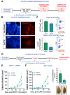
Efficacy of Ad.P60 in experimental breast cancer models. (A) C57Bl/6 mice were subcutaneously injected with 200,000 EO771 cells. When tumor volume reached 500 mm3, mice were treated i.t. by inoculating 6.3 × 10⁷ PFU of Ad.dT or Ad.P60. (B) After 3 days, transduction efficiency was evaluated in tumor cryostat slices by detection of the reporter protein dTomato by fluorescence microscopy. The nuclei of the cells were stained with DAPI. (C) A total of 8 days after inoculation, spleen and tumor-infiltrating Treg populations were evaluated in C57Bl/6J mice transgenic for Foxp3-GFP. Immune populations were analyzed by flow cytometry, studying the presence of CD4+ Foxp3-GFP+ T cells (Student’s t test; * p < 0.05). (D) Tumor growth was evaluated in C57Bl/6 mice bearing syngeneic EO771 tumors (E) and BALB/c mice bearing LM3 tumors (F) that were treated with 3 i.t. injections of Ad.dT or Ad.P60. * p < 0.05; multiple regression analysis. (F) Total number of spontaneous lung metastases in each mouse bearing LM3 tumors. Representative images of lungs are shown, * p < 0.05 (Student’s t test).

Similar articles

Cited by

References

    1. Romano M., Fanelli G., Albany C.J., Giganti G., Lombardi G. Past, Present, and Future of Regulatory T Cell Therapy in Transplantation and Autoimmunity. Front. Immunol. 2019;10:43. doi: 10.3389/fimmu.2019.00043. - DOI - PMC - PubMed
    1. Tanaka A., Sakaguchi S. Targeting Treg cells in cancer immunotherapy. Eur. J. Immunol. 2019;49:1140–1146. doi: 10.1002/eji.201847659. - DOI - PubMed
    1. Colamatteo A., Carbone F., Bruzzaniti S., Galgani M., Fusco C., Maniscalco G.T., Di Rella F., de Candia P., De Rosa V. Molecular Mechanisms Controlling Foxp3 Expression in Health and Autoimmunity: From Epigenetic to Post-translational Regulation. Front. Immunol. 2019;10:3136. doi: 10.3389/fimmu.2019.03136. - DOI - PMC - PubMed
    1. Kaminskiy Y., Kuznetsova V., Kudriaeva A., Zmievskaya E., Bulatov E. Neglected, yet significant role of FOXP1 in T-cell quiescence, differentiation and exhaustion. Front. Immunol. 2022;13:971045. doi: 10.3389/fimmu.2022.971045. - DOI - PMC - PubMed
    1. Wang J., Gong R., Zhao C., Lei K., Sun X., Ren H. Human FOXP3 and tumour microenvironment. Immunology. 2023;168:248–255. doi: 10.1111/imm.13520. - DOI - PubMed

Publication types