

One-Step Assembly of a PRRSV Infectious cDNA Clone and a Convenient CRISPR/Cas9-Based Gene-Editing Technology for Manipulation of PRRSV Genome
- PMID: 37766223
- PMCID: PMC10536534
- DOI: 10.3390/v15091816
One-Step Assembly of a PRRSV Infectious cDNA Clone and a Convenient CRISPR/Cas9-Based Gene-Editing Technology for Manipulation of PRRSV Genome
Abstract
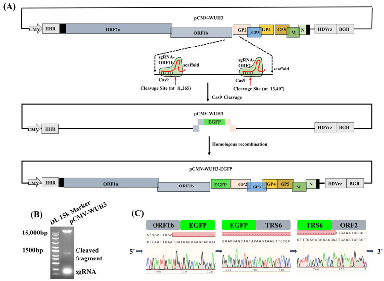
Porcine reproductive and respiratory syndrome (PRRS) has been a persistent challenge for the swine industry for over three decades due to the lack of effective treatments and vaccines. Reverse genetics systems have been extensively employed to build rapid drug screening platforms and develop genetically engineered vaccines. Herein, we rescued recombinant PRRS virus (rPRRSV) WUH3 using an infectious cDNA clone of PRRSV WUH3 acquired through a BstXI-based one-step-assembly approach. The rPRRSV WUH3 and its parental PRRSV WUH3 share similar plaque sizes and multiple-step growth curves. Previously, gene-editing of viral genomes depends on appropriate restrictive endonucleases, which are arduous to select in some specific viral genes. Thus, we developed a restrictive endonucleases-free method based on CRISPR/Cas9 to edit the PRRSV genome. Using this method, we successfully inserted the exogenous gene (EGFP gene as an example) into the interval between ORF1b and ORF2a of the PRRSV genome to generate rPRRSV WUH3-EGFP, or precisely mutated the lysine (K) at position 150 of PRRSV nsp1α to glutamine (Q) to acquire rPRRSV WUH3 nsp1α-K150Q. Taken together, our study provides a rapid and convenient method for the development of genetically engineered vaccines against PRRSV and the study on the functions of PRRSV genes.
Keywords: CRISPR/Cas9; PRRSV; gene-editing; reverse genetics system.
Conflict of interest statement
The authors declare no conflict of interest.
Figures


Similar articles
-
Porcine reproductive and respiratory syndrome virus expressing E2 of classical swine fever virus protects pigs from a lethal challenge of highly-pathogenic PRRSV and CSFV.Vaccine. 2018 May 31;36(23):3269-3277. doi: 10.1016/j.vaccine.2018.04.079. Epub 2018 Apr 30. Vaccine. 2018. PMID: 29724508
-
Evaluation of immune efficacy of recombinant PRRSV vectored vaccine rPRRSV-E2 in piglets with maternal derived antibodies.Vet Microbiol. 2020 Sep;248:108833. doi: 10.1016/j.vetmic.2020.108833. Epub 2020 Aug 27. Vet Microbiol. 2020. PMID: 32891948
-
One-Step Assembly of a Porcine Epidemic Diarrhea Virus Infectious cDNA Clone by Homologous Recombination in Yeast: Rapid Manipulation of Viral Genome With CRISPR/Cas9 Gene-Editing Technology.Front Microbiol. 2022 Feb 10;13:787739. doi: 10.3389/fmicb.2022.787739. eCollection 2022. Front Microbiol. 2022. PMID: 35222326 Free PMC article.
-
Live porcine reproductive and respiratory syndrome virus vaccines: Current status and future direction.Vaccine. 2015 Aug 7;33(33):4069-80. doi: 10.1016/j.vaccine.2015.06.092. Epub 2015 Jul 4. Vaccine. 2015. PMID: 26148878 Review.
-
Engineering the PRRS virus genome: updates and perspectives.Vet Microbiol. 2014 Dec 5;174(3-4):279-295. doi: 10.1016/j.vetmic.2014.10.007. Epub 2014 Oct 23. Vet Microbiol. 2014. PMID: 25458419 Free PMC article. Review.
References
-
- Nelson E.A., Christopher-Hennings J., Drew T., Wensvoort G., Collins J.E., Benfield D.A. Differentiation of U.S. and European isolates of porcine reproductive and respiratory syndrome virus by monoclonal antibodies. J. Clin. Microbiol. 1993;31:5. doi: 10.1128/jcm.31.12.3184-3189.1993. - DOI - PMC - PubMed
Grants and funding
LinkOut - more resources
Full Text Sources
