. 2023 Aug 31;15(9):1855.
doi: 10.3390/v15091855.

An Integrated Research-Clinical BSL-2 Platform for a Live SARS-CoV-2 Neutralization Assay

Affiliations

An Integrated Research-Clinical BSL-2 Platform for a Live SARS-CoV-2 Neutralization Assay

Jing Zou et al. Viruses. .

Abstract

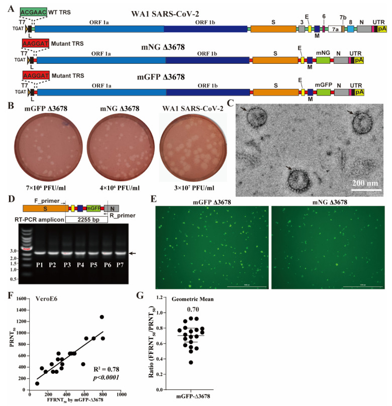
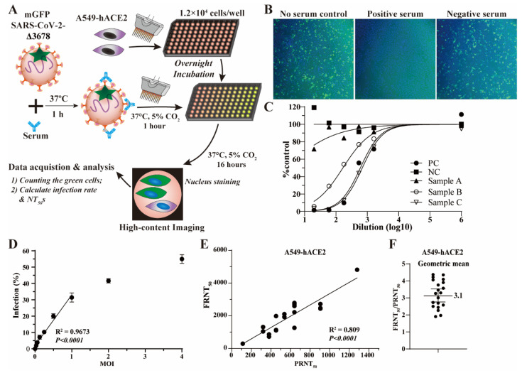
A reliable and efficient serological test is crucial for monitoring neutralizing antibodies against SARS-CoV-2 and its variants of concern (VOCs). Here, we present an integrated research-clinical platform for a live SARS-CoV-2 neutralization assay, utilizing highly attenuated SARS-CoV-2 (Δ3678_WA1-spike). This strain contains mutations in viral transcription regulation sequences and deletion in the open-reading-frames 3, 6, 7, and 8, allowing for safe handling in biosafety level 2 (BSL-2) laboratories. Building on this backbone, we constructed a genetically stable reporter virus (mGFP Δ3678_WA1-spike) by incorporating a modified green fluorescent protein sequence (mGFP). We also constructed mGFP Δ3678_BA.5-spike and mGFP Δ3678_XBB.1.5-spike by substituting the WA1 spike with variants BA.5 and XBB.1.5 spike, respectively. All three viruses exhibit robust fluorescent signals in infected cells and neutralization titers in an optimized fluorescence reduction neutralization assay that highly correlates with a conventional plaque reduction assay. Furthermore, we established that a streamlined robot-aided Bench-to-Clinics COVID-19 Neutralization Test workflow demonstrated remarkably sensitive, specific, reproducible, and accurate characteristics, allowing the assessment of neutralization titers against SARS-CoV-2 variants within 24 h after sample receiving. Overall, our innovative approach provides a valuable avenue for large-scale testing of clinical samples against SARS-CoV-2 and VOCs at BSL-2, supporting pandemic preparedness and response strategies.

Keywords: COVID-19; SARS-CoV-2; high throughput; live attenuated; neutralization assay; serological diagnosis; variants of concern.


Conflict of interest statement

X.X. and P.-Y.S. have filed a patent on the live-attenuated SARS-CoV-2. The other authors declare no conflict of interest.

Figures

Figure 1
Figure 1
mGFP Δ3678 for neutralization assay. (A) Diagram of the construction of mGFP Δ3678. The wild-type and mutated TRSs are colored green and red, respectively. mNG, mNeonGreen; mGFP, sequence-optimized green fluorescence protein. (B) Plaque morphologies on VeroE6-TMPRSS2 cells. Numeric values indicate viral titers of P1 stock. (C) Representative negative-stained electron microscopy micrographs of mGFP Δ3678. Scale bar, 200 nanometers; arrows, virions. (D) Gel analysis. A replicon between F_primer and R_primer was amplified with RT-PCR from the P1-P7 passaged mGFP Δ3678 and analyzed on a 0.6% native agarose gel. DNA ladders are indicated. The arrow indicates the fragment containing the mGFP sequence. (E) Fluorescent focus on VeroE6 cells after 20 h of infection with mGFP Δ3678 or mNG Δ3678. (F) Correlation of mGFP Δ3678 FFRNT with PRNT. PRNT50 was obtained with plaque assay using strain USA-WA1/2020 at BSL-3. The coefficient and p-value (two-tailed) calculated from a linear regression model are shown. (G) Ratios of mGFP Δ3678 FFRNT50 to PRNT50. The geometric mean is shown. The error bar indicates the 95% confidence interval of the geometric mean. The FFRNTs using mGFP Δ3678 and mNG Δ3678 [16] were performed in parallel by using the same set of serum samples.
Figure 2
Figure 2
mGFP Δ3678-based FRNT. (A) Flow chart of mGFP Δ3678 FRNT on A549-hACE2 cells. (B) Representative fluorescent images after 16 h of infection. mGFP Δ3678-infected cells are shown in green. Blue indicates the nucleus. (C) Representative neutralization curve. PC, positive control; NC, negative control. (D) Infection rates at various multiplicities of infection (MOI). The coefficient and two-tailed p-value calculated from the linear regression model are shown. (E) Correlation of mGFP Δ3678 FRNT50 with PRNT50. PRNT50 was obtained with plaque assay using USA-WA1/2020. The coefficient and two-tailed p-value calculated from the linear regression model are shown. (F) Ratios of FFRNT50 to PRNT50. The geometric mean is shown. The error bar indicates the 95% confidence interval of the geometric mean.
Figure 3
Figure 3
B2CNT for testing against SARS-CoV-2 variants BA.5 and XBB.1.5 at BSL-2. (A) Diagram of Bench-to-Clinics Neutralization Test (B2CNT) workflow. The schematic workflow was created with BioRender.com. (B) Infection rates after 16 h of infection with mGFP Δ3678_BA.5-spike or mGFP Δ3678_XBB.1.5-spike. (C) Analysis of the assay robustness. SD, standard deviations; Z’ > 0.5, suitable for high-throughput screening. (D) Correlation of mGFP Δ3678_BA.5-spike FRNT50 with PRNT50. PRNT50 was obtained with plaque assay using the USA-WA1/2020 containing BA.5-spike. The coefficient and p-values (two-tailed) calculated from a linear regression model are shown. (E) Ratios of FFRNT50 using mGFP Δ3678_BA.5-spike to PRNT50. The geometric mean is shown. The error bar indicates the 95% confidence interval of the geometric mean. (F) Correlation of mGFP Δ3678_XBB.1.5-spike FRNT50 with PRNT50. The coefficient and p-values (two-tailed) calculated from a linear regression model are shown. (G) Ratios of FFRNT50 of mGFP Δ3678_XBB.1.5-spike to PRNT50. The geometric mean is shown. The error bar indicates the 95% confidence interval of the geometric mean.

References

    1. Andrews N., Stowe J., Kirsebom F., Toffa S., Rickeard T., Gallagher E., Gower C., Kall M., Groves N., O’Connell A.M., et al. Covid-19 Vaccine Effectiveness against the Omicron (B.1.1.529) Variant. N. Engl. J. Med. 2022;386:1532–1546. doi: 10.1056/NEJMoa2119451. - DOI - PMC - PubMed
    1. Goldberg Y., Mandel M., Bar-On Y.M., Bodenheimer O., Freedman L.S., Ash N., Alroy-Preis S., Huppert A., Milo R. Protection and Waning of Natural and Hybrid Immunity to SARS-CoV-2. N. Engl. J. Med. 2022;386:2201–2212. doi: 10.1056/NEJMoa2118946. - DOI - PMC - PubMed
    1. Grzelak L., Temmam S., Planchais C., Demeret C., Tondeur L., Huon C., Guivel-Benhassine F., Staropoli I., Chazal M., Dufloo J., et al. A comparison of four serological assays for detecting anti-SARS-CoV-2 antibodies in human serum samples from different populations. Sci. Transl. Med. 2020;12:eabc3103. doi: 10.1126/scitranslmed.abc3103. - DOI - PMC - PubMed
    1. Amanat F., White K.M., Miorin L., Strohmeier S., McMahon M., Meade P., Liu W.C., Albrecht R.A., Simon V., Martinez-Sobrido L., et al. An In Vitro Microneutralization Assay for SARS-CoV-2 Serology and Drug Screening. Curr. Protoc. Microbiol. 2020;58:e108. doi: 10.1002/cpmc.108. - DOI - PMC - PubMed
    1. Bewley K.R., Coombes N.S., Gagnon L., McInroy L., Baker N., Shaik I., St-Jean J.R., St-Amant N., Buttigieg K.R., Humphries H.E., et al. Quantification of SARS-CoV-2 neutralizing antibody by wild-type plaque reduction neutralization, microneutralization and pseudotyped virus neutralization assays. Nat. Protoc. 2021;16:3114–3140. - PubMed

LinkOut - more resources