. 2023 Sep 4;15(9):1874.
doi: 10.3390/v15091874.

HDAC-Specific Inhibitors Induce the Release of Porcine Epidemic Diarrhea Virus via the COPII-Coated Vesicles

Affiliations

HDAC-Specific Inhibitors Induce the Release of Porcine Epidemic Diarrhea Virus via the COPII-Coated Vesicles

Ying Yang et al. Viruses. .

Abstract

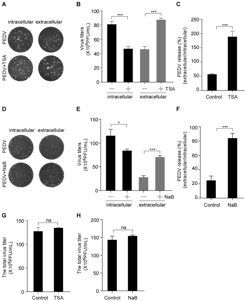
Porcine epidemic diarrhea virus (PEDV) is an alpha-coronavirus causing acute diarrhea and high mortality in neonatal suckling piglets, resulting in huge economic losses for the global swine industry. The replication, assembly and cell egression of PEDV, an enveloped RNA virus, are mediated via altered intracellular trafficking. The underlying mechanisms of PEDV secretion are poorly understood. In this study, we found that the histone deacetylase (HDAC)-specific inhibitors, trichostatin A (TSA) and sodium butyrate (NaB), facilitate the secretion of infectious PEDV particles without interfering with its assembly. We found that PEDV N protein and its replicative intermediate dsRNA colocalize with coat protein complex II (COPII)-coated vesicles. We also showed that the colocalization of PEDV and COPII is enhanced by the HDAC-specific inhibitors. In addition, ultrastructural analysis revealed that the HDAC-specific inhibitors promote COPII-coated vesicles carrying PEDV virions and the secretion of COPII-coated vesicles. Consistently, HDAC-specific inhibitors-induced PEDV particle secretion was abolished by Sec24B knockdown, implying that the HDAC-specific inhibitors-mediated COPII-coated vesicles are required for PEDV secretion. Taken together, our findings provide initial evidence suggesting that PEDV virions can assemble in the endoplasmic reticulum (ER) and bud off from the ER in the COPII-coated vesicles. HDAC-specific inhibitors promote PEDV release by hijacking the COPII-coated vesicles.

Keywords: COPII-coated vesicles; HDAC-specific inhibitors; porcine epidemic diarrhea virus; viral release.


Conflict of interest statement

The authors declare no conflict of interest.

Figures

Figure 1
Figure 1
HDAC-specific inhibitors promote the secretion of PEDV virus particles into the extracellular compartment. (A,B) Determination of the cell viability following treatment with TSA and NaB via CCK-8 test. Vero-E6 cells were pretreated with or without TSA (40 ng/mL) and NaB (2 mM) for 2 h, and then mock-infected or infected with HLJBY (MOI = 0.1). After HLJBY adsorption for 1 h. The cells were further cultured in fresh medium in the presence of TSA and NaB at 12 or 24 h. The infected cell lysates were prepared, and AC-H3, intracellular N, and actin were detected by Western-blot (C,F). The culture medium was divided into two parts; one part was analyzed by Western blotting of extracellular N (C,F), and the other part was titrated by plaque formation assay to measure the extracellular PEDV virus titers (D,G). Graphs show changes in virus titers (E,H); 12 h (×105), 24 h (×106). Student’s t test was used for statistical analysis. **, p < 0.01; ***, p < 0.001. The error bars indicate standard deviation from three independent experiments. In; intracellular, Ex; extracellular.
Figure 2
Figure 2
HDAC-specific inhibitors facilitate the secretion of infectious PEDV particles. Vero-E6 cells were pretreated with or without TSA (40 ng/mL) and NaB (2 mM) for 2 h, and then mock-infected or infected with HLJBY (MOI = 0.1). After HLJBY adsorption for 1 h, the cells were further cultured in fresh medium in the presence of TSA and NaB at 12 or 24 h. The culture medium was first collected, and the infected cells were washed thrice with phosphate-buffered saline (PBS) before adding fresh medium, and then lysed via three freeze/thaw cycles to obtain the intracellular PEDV. (A,D) The virus titers (intracellular and extracellular) were detected by plaque formation assay. (B,E) Graphs show changes in virus titers (intracellular and extracellular); 12 h (×105), 24 h (×106). (C,F) Graphs show PEDV release (ratio of extracellular to intracellular virus titers). (G,H) Graphs show changes in total virus titer. Student’s t test was used for statistical analysis. The error bars indicate standard deviation from three independent experiments. ns, p > 0.05; *, p < 0.05; ***, p < 0.001.
Figure 3
Figure 3
PEDV N protein and its replicative intermediate dsRNA colocalize with COPII-coated vesicles. Vero-E6 cells infected with HLJBY (MOI = 0.1) for 0, 6, 12 and 24 h. The cells were fixed and stained with Sec21p (γ subunit of COPI) antibody (A); COPII antibody (B); clathrin antibody (C); and Alexa 488-conjugated goat anti-rabbit IgG antibodies (green) and then stained with PEDV-N antibody and Alexa 555-conjugated goat anti-mouse IgG antibody (red). The nuclei were stained with DAPI (blue). Images were acquired with a Nikon confocal microscopy. Bottom panel shows a magnified view of the boxed area in panels (AC) (Merged). For quantitative colocalization analysis (QCA), Pearson correlation coefficient (PCC) values were calculated and represent the mean ± SD. (D) Vero-E6 cells infected with HLJBY (MOI = 0.1) for 24 h. The cells were fixed and stained with COPII antibody and Alexa 488-conjugated goat anti-rabbit IgG anti-bodies (green), and then stained with dsRNA antibody and Alexa 555-conjugated goat anti-mouse IgG antibody (red). The nuclei were stained with DAPI (blue). Images were acquired with a Nikon confocal microscopy. Bottom panel shows a magnified view of the boxed area in panel (D) (Merge). For quantitative colocalization analysis (QCA), PCC values were calculated and represent the mean ± SD.
Figure 4
Figure 4
The colocalization between PEDV N and COPII is enhanced by HDAC-specific inhibitors. Vero-E6 cells were pretreated with or without TSA (60 ng/mL) and NaB (4 mM) for 2 h, and then infected with HLJBY (MOI = 1) for 1 h. The cells were further cultured in fresh medium in the presence of TSA and NaB at 8 h. (A) The cells were fixed and stained with COPII antibody and Alexa 488-conjugated goat anti-rabbit IgG antibodies (green) and then stained with PEDV-N antibody and Alexa 555-conjugated goat anti-mouse IgG antibody (red). The nuclei were stained with DAPI (blue). The images were acquired with a Nikon confocal microscopy. (B) Graphs show the PCC of COPII and PEDV N. For quantitative colocalization analysis (QCA), PCC values were calculated and represent the mean ± SD. Student’s t test was used for statistical analysis. ***, p < 0.001; ****, p < 0.0001.
Figure 5
Figure 5
PEDV virions localized to COPII-coated vesicles following HDAC-specific inhibitors treatment. Vero-E6 cells were pretreated with or without TSA (60 ng/mL) and NaB (4 mM) for 2 h, and then mock infected or infected with HLJBY (MOI = 0.1). After HLJBY adsorption for 1 h, the cells were further cultured in fresh medium in the presence of TSA and NaB at 12 or 24 h. (A,C) Virus particles were detected with a transmission electron microscopy (TEM). Arrows indicate transport vesicles containing virions. (B,D) Graphs show the percentage of vesicles carrying PEDV virions. Student’s t test was used for statistical analysis. ***, p < 0.001. The error bars indicate standard deviation from three independent experiments. (E) Detection of COPII-coated vesicles by immunoelectron microscopy (IEM). The red box shows the PEDV virions enclosed in COPII-positive vesicles. The black arrow points to gold nanoparticles (10 nm)-labeled COPII. The white arrow points to PEDV virions.
Figure 6
Figure 6
Suppression of HDAC-specific inhibitors-induced PEDV secretion by Sec24B knockdown. (A) Schematic diagram of the vesicle budding process in the COPII complex. Vero-E6 cells were transfected with nontargeting scrambled siRNA or targeting Sec23A/Sec24B siRNA for 48 h, and then pretreated with or without TSA (40 ng/mL) for 2 h, followed by HLJBY (MOI = 0.1) infection. After HLJBY adsorption for 1 h, the cells were further cultured in fresh medium in the presence of TSA for 12 h. (B) Sec23A/Sec24B and actin were detected by immunoblotting. (C,D) The cells were fixed and stained with Sec24B or COPII antibody and Alexa 488-conjugated goat anti-rabbit IgG antibodies (green) and then stained with PEDV-N antibody and Alexa 555-conjugated goat anti-mouse IgG antibody (red). The nuclei were stained with DAPI (blue). The images were acquired with a Nikon immunofluorescence microscopy. (E) Virus titers were measured via plaque formation assay, and the graphs show changes in virus titers (intracellular and extracellular); 12 h (×105). (F) Graph shows PEDV release (ratio of extracellular to intracellular virus titers). Student’s t test was used for statistical analysis. ***, p < 0.001; ****, p < 0.0001. The error bars indicate standard deviation from three independent experiments.
Figure 7
Figure 7
The interplay between HDAC-specific inhibitors and COPII-coated vesicles during PEDV infection. Following the entry of PEDV into host cells and replication in the cytoplasm, the newly synthesized PEDV viral components and PEDV virus particles are transported to Golgi apparatus by the COPII-coated vesicles. HDAC-specific inhibitors facilitate PEDV release by promoting the secretion of COPII-coated vesicles carrying PEDV virions.

Similar articles

Cited by

References

    1. Pillaiyar T., Meenakshisundaram S., Manickam M. Recent discovery and development of inhibitors targeting coronaviruses. Drug Discov. Today. 2020;25:668–688. doi: 10.1016/j.drudis.2020.01.015. - DOI - PMC - PubMed
    1. Schaeffer R., Temeeyasen G., Hause B.M. Alphacoronaviruses Are Common in Bats in the Upper Midwestern United States. Viruses. 2022;14:184. doi: 10.3390/v14020184. - DOI - PMC - PubMed
    1. Sun L., Chen H., Ming X., Bo Z., Shin H.J., Jung Y.S., Qian Y. Porcine Epidemic Diarrhea Virus Infection Induces Caspase-8-Mediated G3BP1 Cleavage and Subverts Stress Granules To Promote Viral Replication. J. Virol. 2021;95:e02344-20. doi: 10.1128/JVI.02344-20. - DOI - PMC - PubMed
    1. Hasöksüz M., Kiliç S., Saraç F. Coronaviruses and SARS-COV-2. Turk. J. Med. Sci. 2020;50:549–556. doi: 10.3906/sag-2004-127. - DOI - PMC - PubMed
    1. Jung K., Saif L.J., Wang Q. Porcine epidemic diarrhea virus (PEDV): An update on etiology, transmission, pathogenesis, and prevention and control. Virus. Res. 2020;286:198045. doi: 10.1016/j.virusres.2020.198045. - DOI - PMC - PubMed