. 2023 Sep 5;15(9):1883.
doi: 10.3390/v15091883.

Comparison of Gross Pathology between Classical and Recombinant Lumpy Skin Disease Viruses

Affiliations

Comparison of Gross Pathology between Classical and Recombinant Lumpy Skin Disease Viruses

Irina Shumilova et al. Viruses. .

Abstract

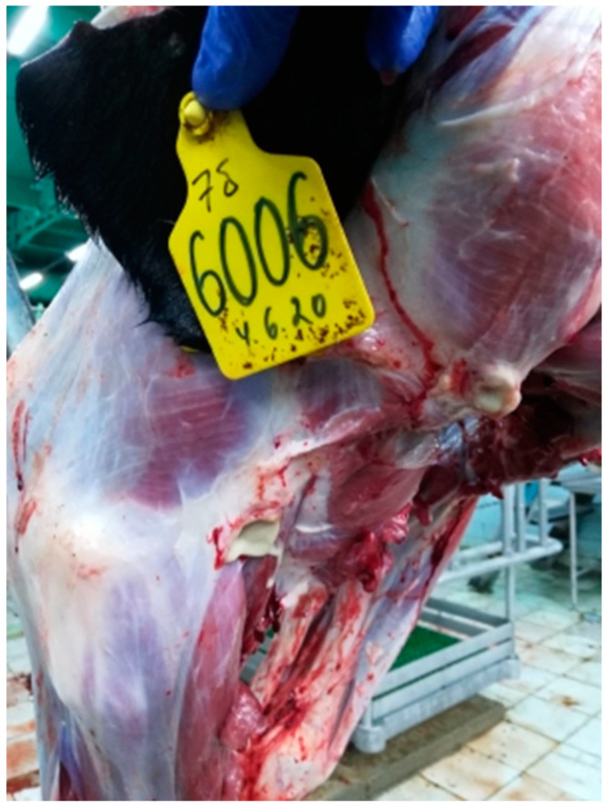
The pathology caused by three different isolates of lumpy skin disease virus, classical field cluster 1.2 strain Dagestan/2015, recombinant vaccine-like cluster 2.1 strain Saratov/2017, and cluster 2.2 strain Udmurtiya/2019, in cattle was compared from experimental infections. The infection of cattle was performed using intravenous administration of 2 mL of 105 TCID50/mL of each specific LSDV. Both classical and recombinant forms of LSDV cause pathological changes in the skin and lymph nodes, as well as the trachea and lungs. Due to circulatory disorders in the affected organs, multiple areas of tissue necrosis were observed, which, with the resurgence of secondary microflora, led to the development of purulent inflammation. Observed pathological changes caused by the recombinant vaccine-like strain Udmurtiya/2019 were characterized by a more pronounced manifestation of the pathoanatomical picture compared to the classical field strains Dagestan/2015 and Saratov/2017. Interestingly, Dagestan/2015 and Udmurtiya/2019 caused damage to the lymph nodes, characterized by serous inflammation and focal purulent lymphadenitis caused by purulent microflora. "Saratov/2017" did not cause pathology in the lymph nodes. All LSDVs were virulent and caused pathology, which was not distinguishable between viruses. This data set will serve as the experimentally validated basis for the comparative examination of novel LSDV strains in gross pathology.

Keywords: experimental bioassays; lumpy skin disease virus; lumpy skin diseases; necropsy; pathology; recombinant virus.


Conflict of interest statement

The authors declare that the research was conducted in the absence of any commercial or financial relationships that could be construed as a potential conflict of interest.

Figures

Figure 1
Figure 1
Pathological changes in skin, subcutaneous, and muscle tissues in cattle infected with LSDV Dagestan/2015. (A) Multiple focal necrotic dermatomyositis. (B) Focal and fine-spotted hemorrhages in subcutaneous tissue. Subcutaneous tissue is moist and shiny, with multiple jagged-edged red foci of various shapes and sizes on the periphery; local, small hemorrhages. Areas with affected outer layers of muscle were observed as red-colored muscle lesions of irregular sizes and shapes.
Figure 2
Figure 2
Pathological changes in skin, subcutaneous, and muscle tissue in cattle caused by LSDV Udmurtiya/2019. (A) Multiple nodular dermatitis on the skin surface; multiple swollen, dense, rounded-shaped foci of various sizes. In some cases, these areas had raised edges and a concavity in the central part, which is characterized by a rough surface and grayish-red color. (B) Multiple focal purulent-necrotic dermatitis (inner surface of the skin). (C) Multiple spot and fine-spotted hemorrhages in the subcutaneous tissue; the subcutaneous tissue is wet and shiny, with multiple small-spotted hemorrhages and focal lesions of various shapes and sizes, with uneven edges, purplish-red in color, sometimes with signs of purulent melting.
Figure 3
Figure 3
Pathological changes in skin, subcutaneous, and muscle tissues in cattle caused by LSDV Saratov/2017. Focal necrotic dermatitis. Multiple spot hemorrhages in the subcutaneous tissue. Subcutaneous tissue is moist and shiny, has focal lesions of red color, is rounded or irregular in shape, has uneven edges, and has a structureless central part of red-cyanotic color.
Figure 4
Figure 4
Multiple erosive and ulcerative dermatitis of the nasal mirror skin. (A)—Bull infected with Dagestan/2015. The skin of the nasal mirror is moist and shiny, and around it there are multiple defects of a rounded or oval shape with smooth edges; (B)—Bull infected with Udmurtiya/2019. Erosive and ulcerative dermatitis of the nasal mirror. On the dorsal surface of the nasal mirror as well as on the inner surface of the skin of the nasal passages, focal lesions of a rounded or oval shape with uneven edges and deepening in the center were found.
Figure 5
Figure 5
Erosive-ulcerative glossitis—the tongue is enlarged in size and edematous. The mucous membrane is swollen, and in the area of the tip of the tongue, it has focal lesions of irregular shape, merging with each other and penetrating deep into the tissue. On the mucous membrane in the area of the body and the root of the tongue, there are multiple lesions of a rounded shape with towering edges and a sinking center, the surface of which is uneven and grayish-pink in color.
Figure 6
Figure 6
Croup pneumonia complicated by an abscess. Dagestan/2015. (A) The lungs are enlarged, bumpy from the surface, and areas of dense consistency are revealed; (B) On the incision—moist, the interstitial is expanded, the affected areas are dark red, and, in places, there are cavities filled with purulent contents.
Figure 7
Figure 7
Croup pneumonia by Saratov/2017. (A,B) Correspond to animals infected intravenously, while (C,D) are in-contact control animals. (A)—Croup pneumonia in animals infected intravenously; (B)—The lungs are enlarged, lumpy, and the lung tissue has collapsed in places along the edges; on the incision—grayish-red, the interstitial is expanded; (C)—Croup pneumonia by Saratov/2017 with foci of necrosis (in contact animal); (D)—Croup pneumonia with foci of necrosis. Some lobes of the lung are enlarged, dark red in color, and dense in consistency. In some places, there are foci of reddish-bluish color protruding above the surface of the lungs and containing structureless grayish-yellowish inclusions (in contact animals).
Figure 8
Figure 8
Croup pneumonia. Udmurtiya/2019. The lungs are enlarged in size, bumpy, dense in consistency, and red-bluish in color. The interstitium is expanded and gray-white in color.
Figure 9
Figure 9
Acute serous tracheitis with multiple spots and small-spotted hemorrhages. Dagestan/2015. The mucous membrane of the trachea is swollen, grayish-pink in color, and, in places, has intense red staining with multiple small hemorrhages.
Figure 10
Figure 10
Acute catarrhal tracheitis with single spot and small-spotted hemorrhages. Udmurtiya/2019. The mucous membrane of the trachea is grayish-red in places with a bluish tinge; moist, shiny, straw-colored mucus is noted, as well as spot and fine-spotted hemorrhages. On the mucous membrane in the upper third of the trachea, a large oval-shaped, gray-red focus is visualized, under which the submucosal base is impregnated with a reddish liquid.
Figure 11
Figure 11
Serous lymphadenitis of the pharyngeal lymph nodes with spot hemorrhages. Dagestan/2015. Lymph nodes are enlarged, juicy, gray-white in color, and have spot hemorrhages.
Figure 12
Figure 12
Serous lymphadenitis. Udmurtiya/2019. The lymph node is enlarged in size, moist, shiny, and reddish-brown in color on the incision.
Figure 13
Figure 13
Focal purulent lymphadenitis with Udmurtiya/2019. The lymph node is enlarged, with round and oval foci filled with purulent contents.

References

    1. Sprygin A., Sainnokhoi T., Gombo-Ochir D., Tserenchimed T., Tsolmon A., Byadovskaya O., Ankhanbaatar U., Mazloum A., Korennoy F., Chvala I. Genetic characterization and epidemiological analysis of the first lumpy skin disease virus outbreak in Mongolia, 2021. Transbound. Emerg. Dis. 2022;69:3664–3672. doi: 10.1111/tbed.14736. - DOI - PubMed
    1. Fagbo S., Coetzer J.A.W., Venter E.H. Seroprevalence of Rift Valley fever and lumpy skin disease in African buffalo (Syncerus caffer) in the Kruger National Park and Hluhluwe-iMfolozi Park, South Africa. J. S. Afr. Vet. Assoc. 2014;85:e1–e7. doi: 10.4102/jsava.v85i1.1075. - DOI - PubMed
    1. Le Goff C., Lamien C.E., Fakhfakh E., Chadeyras A., Aba-Adulugba E., Libeau G., Tuppurainen E., Wallace D.B., Adam T., Silber R., et al. Capripoxvirus G-protein-coupled chemokine receptor: A host-range gene suitable for virus animal origin discrimination. J. Gen. Virol. 2009;90:1967–1977. doi: 10.1099/vir.0.010686-0. - DOI - PubMed
    1. Kononov A., Prutnikov P., Shumilova I., Kononova S., Nesterov A., Byadovskaya O., Pestova Y., Diev V., Sprygin A. Determination of lumpy skin disease virus in bovine meat and offal products following experimental infection. Transbound. Emerg. Dis. 2019;66:1332–1340. doi: 10.1111/tbed.13158. - DOI - PubMed
    1. Sprygin A., Pestova Y., Wallace D.B., Tuppurainen E., Kononov A.V. Transmission of lumpy skin disease virus: A short review. Virus Res. 2019;269:197637. doi: 10.1016/j.virusres.2019.05.015. - DOI - PubMed

Publication types

LinkOut - more resources