Review
. 2023 Sep 12;15(9):1913.
doi: 10.3390/v15091913.

Hepatitis B Virus Epsilon (ε) RNA Element: Dynamic Regulator of Viral Replication and Attractive Therapeutic Target

Affiliations
Review

Hepatitis B Virus Epsilon (ε) RNA Element: Dynamic Regulator of Viral Replication and Attractive Therapeutic Target

Lukasz T Olenginski et al. Viruses. .

Abstract

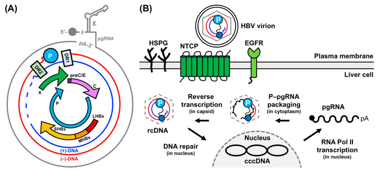
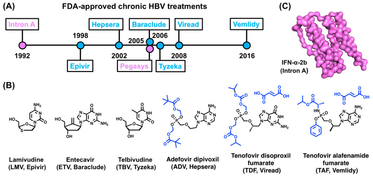
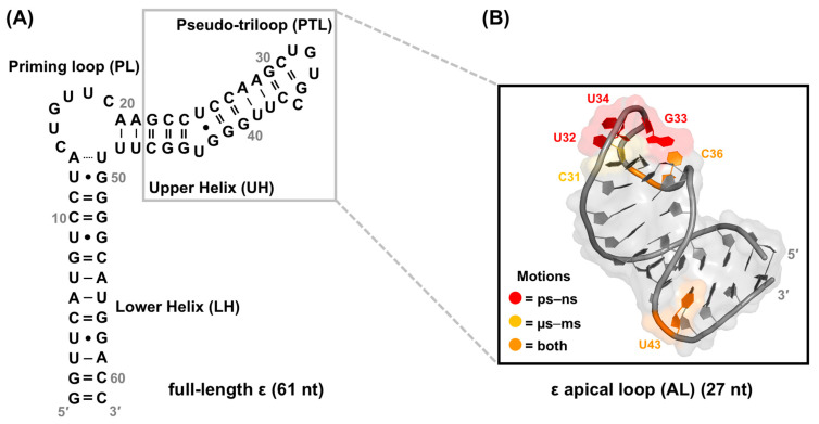
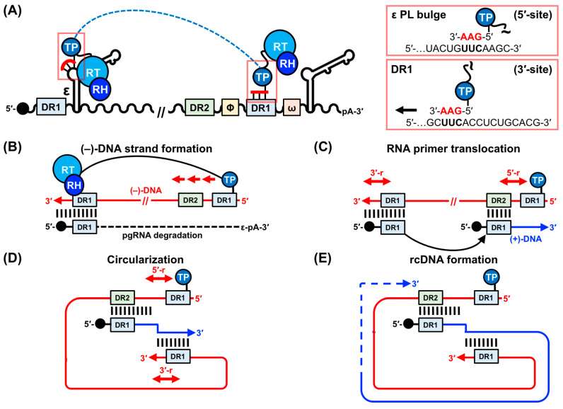
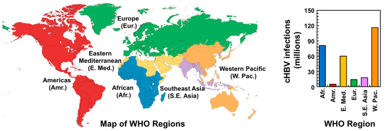
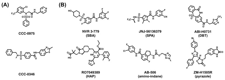
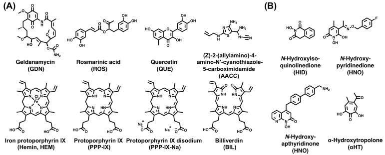
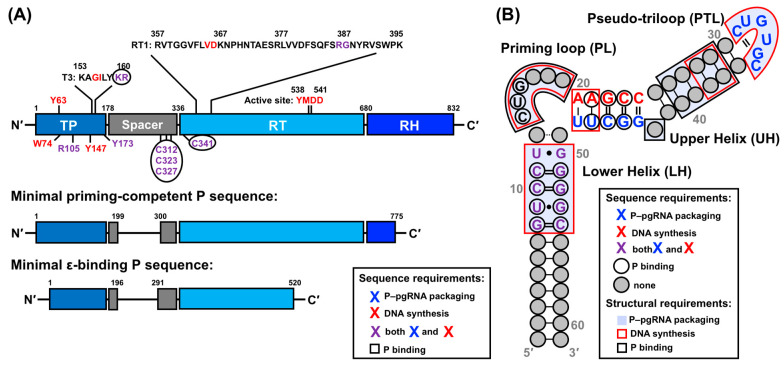
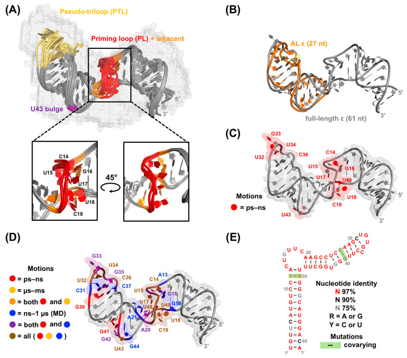
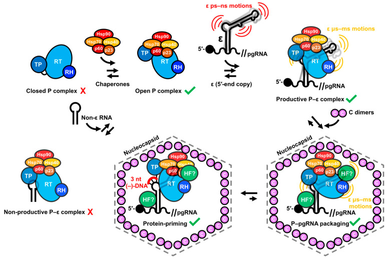
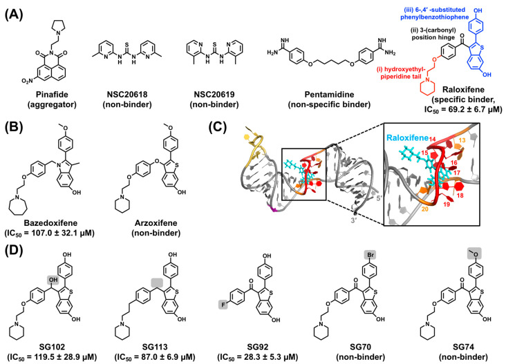
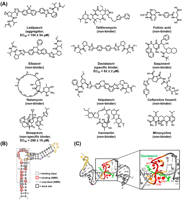
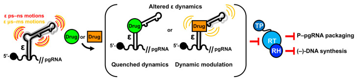
Hepatitis B virus (HBV) chronically infects millions of people worldwide, which underscores the importance of discovering and designing novel anti-HBV therapeutics to complement current treatment strategies. An underexploited but attractive therapeutic target is ε, a cis-acting regulatory stem-loop RNA situated within the HBV pregenomic RNA (pgRNA). The binding of ε to the viral polymerase protein (P) is pivotal, as it triggers the packaging of pgRNA and P, as well as the reverse transcription of the viral genome. Consequently, small molecules capable of disrupting this interaction hold the potential to inhibit the early stages of HBV replication. The rational design of such ligands necessitates high-resolution structural information for the ε-P complex or its individual components. While these data are currently unavailable for P, our recent structural elucidation of ε through solution nuclear magnetic resonance spectroscopy marks a significant advancement in this area. In this review, we provide a brief overview of HBV replication and some of the therapeutic strategies to combat chronic HBV infection. These descriptions are intended to contextualize our recent experimental efforts to characterize ε and identify ε-targeting ligands, with the ultimate goal of developing novel anti-HBV therapeutics.

Keywords: HBV; RNA; small molecules; structure; therapeutics.


Conflict of interest statement

The authors declare no conflict of interest.

Figures

Figure 1
Figure 1
HBV genome organization and lifecycle. (A) Schematic of the 3.2 kB HBV rcDNA genome, depicting the pgRNA, negative (−)- and positive (+)-DNA strands, and its four open reading frames and seven gene products. Critical elements such as the attachment of P to (−)-DNA, direct repeats 1 (DR1) and 2 (DR2), and ε are highlighted. (B) Schematic of HBV genome replication, encompassing liver cell entry to complete genome conversion. This figure is adapted from [29,34].
Figure 4
Figure 4
FDA-approved cHBV treatments. (A) Drug approval timeline. Trade names of NRTIs and IFN-α are shown in blue and purple, respectively. (B) Chemical structures of the NRTI prodrugs from (A), with regions that are removed during activation shown in blue. (C) Structure of IFN-α-2b (PDB 1rh2) [83]. Trade names for drugs in (B,C) are shown in parentheses.
Figure 7
Figure 7
Summary of early NMR studies of ε. (A) Secondary structure of full-length ε [21,22] with abbreviations for structure elements that will be used throughout the text. The dotted line between A13–U49 signifies the absence of unambiguous evidence of base pairing from NMR measurements [28] (B) Top-ranked AL ε solution NMR conformer (PDB 2ixy) [182] with NMR data [183] mapped onto the structure. The AL comprises nucleotides in the UH and PTL, as indicated by the gray box in (A). Nucleotides in the PTL and U43 bulge (based on full-length numbering) exhibit both ps–ns and µs–ms motions, which have been hypothesized to facilitate P binding.
Figure 2
Figure 2
HBV genome replication. (A) The binding of ε to P initiates P–pgRNA packaging and subsequent reverse transcription. Before (−)-DNA strand formation, the TP domain synthesizes a 3 nt DNA (5′-GAA-3′), which is templated by the ε PL bulge (5′-UUC-3′). The TP-linked DNA then translocates (indicated by a dashed blue line) to the 3′-end DR1 motif (5′-UUC-3′) where (−)-DNA strand elongation begins. (B) The TP-linked DNA extends (−)-DNA strand synthesis toward the 5′-end of the pgRNA, which is concurrently degraded by the RH domain of P. (C) The RNA primer subsequently translocates to the DR2 motif and extends toward the 5′-end of the (−)-DNA strand, initiating (+)-DNA strand synthesis. Here, the terms 3′- and 5′-r refer to the 10 nt redundancy that is generated with the (−)-DNA strand. (D) After copying the 5′-r, the growing 3′-end of the (+)-DNA strand translocates to the 5′-r on the (−)-DNA strand to permit further elongation. (E) The final extension of the (−)-DNA strand template yields (+)-DNA strands of various lengths to form the new rcDNA. This figure is adapted from [29].
Figure 3
Figure 3
cHBV infection statistics. Map of the WHO regions (left) and their cHBV infection statistics (right). Data were accessed from the WHO website (https://www.who.int/news-room/fact-sheets/detail/hepatitis-b (accessed on 31 August 2023)).
Figure 5
Figure 5
Small molecules targeting the conversion of HBV rcDNA to cccDNA. Chemical structures of (A) disubstituted sulfonamide cccDNA formation inhibitors and (B) CAMs in recent and ongoing clinical trials from the SBA, SPA, DBT, HAP, amino-indane, and pyrazole chemotypes.
Figure 6
Figure 6
HBV P and ε–P complex targeting small molecules. Chemical structures of (A) ε–P complex inhibitors and (B) RH inhibitors from the HID, HNO, HPD, and αHT chemotypes.
Figure 8
Figure 8
Sequence and structural dependencies of P protein and ε RNA in HBV replication. (A) The sequence and/or structural requirements of P for ε–P binding, specifically for the RT domain. (B) The sequence and/or secondary structure prerequisites of ε for ε–P binding, P–pgRNA packaging, and DNA synthesis. Depictions in (A,B) are based on a synthesis of prior biochemical and mutational studies [19,21,22,25,146,177,178,197].
Figure 9
Figure 9
Summary of the structural dynamics data of full-length ε. (A) Bundle of the top-10 lowest energy ε structures (PDB 6var) [28] generated by Xplor-NIH [199], with an RMSD of 1.8 Å and displayed within the SAXS envelope and with important structural regions colored. A close-up view of the PL in three NMR conformers (rank 3, 5, and 6) is also shown. These conformers share the backbone kink centered at U15, followed by partially stacked G16 and U17. (B) Overlay of the top-ranked full-length ε NMR conformer (PDB 6var) [28] and AL ε NMR conformer (PDB 2ixy) [182]. Structures show strong agreement (RMSD of 1.7 Å). (C) Top-ranked full-length ε solution NMR conformer (PDB 6var) [28] with NMR dynamics data [28] mapped onto the structure. (D) Similar to (C) but from our recent combinined NMR and MD data [198]. (E) Rfam (RF01047) representation [200,201,202] of ε, showing structure and sequence conservation. Nucleotide identity calculations and covarying mutations are based on 36 sequences from six species, including different HBV genotypes but excluding the related avian HBV sequences (Rfam RF01313). As depicted in (CE), highly conserved nucleotides in and adjacent to the PL, PTL, and U43 bulge exhibit motions across multiple timescales.
Figure 10
Figure 10
Schematic of the molecular and biophysical determinants of HBV replication. This model draws on mutational and biochemical data of P and ε [19,21,22,25,146,177,178,197] and our recent NMR structural dynamic studies of ε [28,198]. Additional details can be found in the text.
Figure 11
Figure 11
HTS of full-length ε. (A) Chemical structure of SMM-derived lead compounds with their NMR-derived binding mode (i.e., specific or nonspecific binder, nonbinder, or aggregator) and dye-displacement-derived affinity to full-length ε (measured by IC50) shown in parentheses. (B) Chemical structure of SERMs with their affinity to full-length ε reported as in (A). (C) Top-ranked Raloxifene docking pose to ε R3 (PDB 6var) [28], which is colored as in Figure 9A. Raloxifene is depicted in cyan sticks and interacting nucleotides are labeled. As seen from the close-up view of the binding pocket, the Raloxifene hydroxyethylpiperidine tail does not participate in binding. (D) Chemical structure of Raloxifene analog library with their affinity to full-length ε reported as in (A). Any modification to the three Raloxifene “units” (shown in (A)) is highlighted with a gray box.
Figure 12
Figure 12
VS of full-length ε. (A) Chemical structures of VS-identified lead compounds with their dye-displacement derived binding mode (i.e., specific or nonspecific binder, nonbinder, or aggregator) and affinities to full-length ε (measured by EC50) shown in parentheses. (B). Representative data from dye-displacement, NMR titration, and computational docking analysis are shown mapped onto the secondary structure of ε. Collectively, these data agree that Daclatasvir targets FL ε mainly at its PL and upper segment of the LH. (C) Top-ranked Daclatasvir docking pose to ε R3 (PDB 6var) [28]. Daclatasvir is depicted in green sticks and interacting nucleotides are labeled. Structure representations in (B,C) are colored as in Figure 9A.
Figure 13
Figure 13
Schematic of dynamic-regulating, ε-targeting small molecules. This model draws on our recent NMR structural dynamic studies ε and ligand-bound computational modeling [28,198]. Additional detail can be found in the text.

Similar articles

Cited by

References

    1. Galibert F., Mandart E., Fitoussi F., Tiollais P., Charnay P. Nucleotide sequence of the hepatitis B virus genome (subtype ayw) cloned in E. coli. Nature. 1979;281:646–650. doi: 10.1038/281646a0. - DOI - PubMed
    1. Valenzuela P., Gray P., Quiroga M., Zaldivar J., Goodman H.M., Rutter W.J. Nucleotide sequence of the gene coding for the major protein of hepatitis B virus surface antigen. Nature. 1979;280:815–819. doi: 10.1038/280815a0. - DOI - PubMed
    1. Charnay P., Mandart E., Hampe A., Fitoussi F., Tiollais P., Galibert F. Localization on the viral genome and nucleotide sequence of the gene coding for the two major polypeptides of the Hepatitis B surface antigen (HBs Ag) Nucleic Acids Res. 1979;7:335–346. doi: 10.1093/nar/7.2.335. - DOI - PMC - PubMed
    1. Pasek M., Goto T., Gilbert W., Zink B., Schaller H., Mackay P., Leadbetter G., Murray K. Hepatitis B virus genes and their expression in E. coli. Nature. 1979;282:575–579. doi: 10.1038/282575a0. - DOI - PubMed
    1. Robinson W.S., Clayton D.A., Greenman R.L. DNA of a Human Hepatitis B Virus Candidate. J. Virol. 1974;14:384–391. doi: 10.1128/jvi.14.2.384-391.1974. - DOI - PMC - PubMed

Publication types

LinkOut - more resources