. 2023 Sep 21;15(9):1971.
doi: 10.3390/v15091971.

The Disruption of a Nuclear Export Signal in the C-Terminus of the Herpes Simplex Virus 1 Determinant of Pathogenicity UL24 Protein Leads to a Syncytial Plaque Phenotype

Affiliations

The Disruption of a Nuclear Export Signal in the C-Terminus of the Herpes Simplex Virus 1 Determinant of Pathogenicity UL24 Protein Leads to a Syncytial Plaque Phenotype

Carmen Elena Gonzalez et al. Viruses. .

Abstract

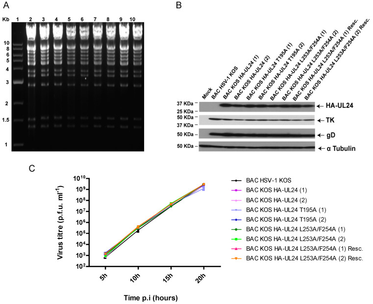
UL24 of herpes simplex virus 1 (HSV-1) has been shown to be a determinant of pathogenesis in mouse models of infection. The N-terminus of UL24 localizes to the nucleus and drives the redistribution of nucleolin and B23. In contrast, when expressed alone, the C-terminal domain of UL24 accumulates in the Golgi apparatus; its importance during infection is unknown. We generated a series of mammalian expression vectors encoding UL24 with nested deletions in the C-terminal domain. Interestingly, enhanced nuclear staining was observed for several UL24-deleted forms in transient transfection assays. The substitution of a threonine phosphorylation site had no effect on UL24 localization or viral titers in cell culture. In contrast, mutations targeting a predicted nuclear export signal (NES) significantly enhanced nuclear localization, indicating that UL24 is able to shuttle between the nucleus and the cytoplasm. Recombinant viruses that encode UL24-harboring substitutions in the NES led to the accumulation of UL24 in the nucleus. Treatment with the CRM-1-specific inhibitor leptomycin B blocked the nuclear export of UL24 in transfected cells but not in the context of infection. Viruses encoding UL24 with NES mutations resulted in a syncytial phenotype, but viral yield was unaffected. These results are consistent with a role for HSV-1 UL24 in late cytoplasmic events in HSV-1 replication.

Keywords: UL24; herpes simplex virus; nuclear export signal; syncytia.


Conflict of interest statement

The authors declare no conflict of interest.

Figures

Figure 1
Figure 1
Deletion mutations targeting the C-terminal domain of HSV-1 UL24: (A) The schematic representation of the deletion series created for the C-terminal domain of UL24. The HA tag is represented with a white box, and the UL24 amino acid sequence is shown with the gray and black boxes to the right. The numbers below the boxes represent UL24 amino acids. The black box represents the amino acids corresponding to the fragment of UL24 that was amplified via PCR to construct the respective plasmid, with the position of the primers used for amplification shown in blue and red. Stop codons were introduced into the three ORFs to block translation after aa 265, 252, 240, 219or 197. The crossed lines in the gray box represent the deleted regions of UL24. (B) Western blot analysis showing the expression of the full-length and truncated HA-UL24 proteins 48 h post-transfection. Cell lysates of COS-7 cells transfected with a plasmid encoding wild-type HA-UL24 or encoding HA-UL24 with the indicated deletion were analyzed using Western blotting with an anti-HA antibody (top panel). The blot was subsequently stripped and incubated with an antibody against α Tubulin, which served as a loading control (bottom panel). The sizes of molecular weight markers are indicated to the left of the panels.
Figure 2
Figure 2
Deletions in the C-terminus of UL24 lead to nuclear sequestration of the protein. COS-7 cells were transiently transfected with plasmids encoding the indicated forms of UL24 protein: (AC) Confocal images showing the localization of wild-type HA-UL24 and the N-terminal and C-terminal domains. (DH) The localization of HA-UL24 variants with C-terminal truncations. The amino acid indicated represents the last one before the inserted stop codon. HA-UL24 was detected via indirect immunofluorescence using a monoclonal antibody directed against HA (green). Nuclei were stained with Draq5 (blue). Merged images are shown in the right-hand panels. Scale bars represent 10 μm.
Figure 3
Figure 3
Site-directed mutagenesis targeting the C-terminal domain of HSV-1 UL24: (A) The primary sequence of the C-terminal portion of UL24 (aa 193–269). Amino acids in red represent the last amino acid expressed for each of the various truncated forms of UL24. The yellow rectangle delineates a putative phosphorylation site (aa 195–197) identified using the MotifScan software. The blue rectangle delineates a possible NES sequence (aa 250–258) identified through comparison with known viral NESs; the positions of the hydrophobic amino acids thought to form the non-classical NES in UL24 are shown below the blue rectangle: Ø represents a hydrophobic residue, and X indicates any residue. Amino acids in green were replaced with alanines to test their importance in the subcellular localization of the protein. (B) Graphic representation showing the position of the phosphorylation site (red arrow) and predicted NES (blue line) in the HSV-1 UL24 full-length protein and truncated versions. The HA tag is represented with a gray box. (C) Western blot analysis showing the expression of the wild-type and substituted forms of HA-UL24 48 h post-transfection. Cell lysates of COS-7 cells transfected with plasmids encoding various forms of HA-UL24 were analyzed via Western blotting with an anti-HA antibody (top panel). The blot was subsequently stripped and incubated with an antibody against α Tubulin, which served as a loading control (bottom panel). The sizes of molecular weight markers are indicated to the left of the panels. Arrows to the right of the panels indicate the positions of HA-UL24 and α Tubulin.
Figure 4
Figure 4
Substitution of hydrophobic residues in the HSV-1 UL24 NES blocks the nuclear export of the protein: (AE) Confocal images show the localization of wild-type HA-UL24 and substituted forms with an altered phosphorylation site or NES sequence in transiently transfected COS-7 cells. HA-UL24 was detected via indirect immunofluorescence using a monoclonal antibody directed against HA (green). Nuclei were stained with Draq5 (blue). Merged images are shown in the right-hand panels. Scale bars represent 10 μm.
Figure 5
Figure 5
The UL24 NES is sensitive to leptomycin B. COS-7 cells were transiently transfected with a plasmid encoding wild-type HA-UL24 (AC) or with plasmids encoding HA-UL24 with an altered phosphorylation site (DF) or substituted NES sequence (GI). Briefly, 22 h post-transfection, cells were treated for 5 h with 10 ng/mL LMB (B,E,H) or with 25 ng/mL LMB (C,F,I). Representative images acquired using confocal microscopy are shown. The subcellular distribution of HA-UL24 was examined using a monoclonal antibody directed against HA and a secondary antibody conjugated with Alexa-488 (green). Nuclei were stained with Draq 5 (blue). Scale bars represent 10 μm. (J,K) The quantification of the nuclear–cytoplasmic (N/C) ratio of the mean intensity of fluorescence for HA-UL24 (J) and HA-UL24 T195A (K) in COS-7 cells in the presence of 10 ng/mL or 25 ng/mL LMB compared with the vehicle alone. The mean N/C ratios were determined from 30 cells over 3 independent experiments. Error bars represent the standard deviation of the mean. **, p < 0.0003 compared with control using one-way ANOVA following a Bonferroni correction of the alpha value for multiple comparisons.
Figure 6
Figure 6
Characterization of recombinant HSV-1 BACs: (A) The EcoRV restriction pattern of genomes of the recombinant BAC HSV-1 strains carrying mutations in UL24. DNA was resolved on a 0.7% agarose gel and stained with ethidium bromide. The numbers (1) and (2) in the names of the viruses represent the different isolates. Lane 1, 1kb DNA ladder; lane 2, BAC HSV-1 KOS; lane 3, BAC_KOS HA-UL24 (1); lane 4, BAC_KOS HA-UL24 (2); lane 5, BAC_KOS HA-UL24 T195A (1); lane 6, BAC_KOS HA-UL24 T195A (2); lane 7, BAC_KOS HA-UL24 L253A/F254A (1); lane 8, BAC_KOS HA-UL24 L253A/F254A (2); lane 9, BAC_KOS HA-UL24 L253A/F254A (1) Resc.; lane 10, BAC_KOS HA-UL24 L253A/F254A (2) Resc. The sizes of the molecular markers are shown on the left. (B) Western blot analysis showing the expression of HA-UL24, HSV-1 TK, and HSV-1 gD by the different recombinant viruses. Lysates of Vero cells mock-infected or infected at an MOI of 10 with the indicated recombinant virus were analyzed via Western blotting with specific antibodies against HA, TK, and gD. The blot was also probed with an antibody against α Tubulin, which served as a loading control. The sizes of molecular weight markers are indicated to the left of the panels. Arrows to the right of the panels indicate the positions of the different proteins. (C) Replication curves for the recombinant viruses. Each virus indicated was used to infect Vero cells in duplicate at an MOI of 5. The supernatant and the cell-associated virus were collected at the indicated time points, and the total infectious virus produced was titrated. Error bars represent the standard error of the mean of two independent experiments.
Figure 7
Figure 7
Mutations targeting the UL24 NES result in syncytial plaques. Representative plaques observed at 37 °C and 39 °C on Vero cells: (A) Plaques produced by the viruses BAC HSV-1 KOS, BAC_KOS HA-UL24 (1), and BAC_KOS HA-UL24 T195A (1) and (2). (B) Plaques produced by the viruses BAC_KOS HA-UL24 L253A/F254A (1) and (2) and BAC_KOS HA-UL24 L253A/F254A (1) Resc and (2) Resc.
Figure 8
Figure 8
Mutations targeting the UL24 NES block nuclear export of UL24 during infection. Vero cells grown on coverslips were infected with the indicated virus at an MOI of 10. At 9 and 18 hpi; the cells were washed, fixed, and immunostained; and representative images were acquired using confocal microscopy. The intracellular distribution of HA-UL24 was examined using a monoclonal antibody directed against HA and a secondary antibody conjugated with Alexa-488. (A) vHA-UL24, BAC_KOS HA-UL24 (1), BAC_KOS HA-UL24 T195A (1), BAC_KOS HA-UL24 T195A (2); (B) BAC_KOS HA-UL24 L253A/F254A (1), BAC_KOS HA-UL24 L253A/F254A (2); BAC_KOS HA-UL24 L253A/F254A (1) Rescue; BAC_KOS HA-UL24 L253A/F254A (2) Rescue. Scale bars represent 10 μm.
Figure 9
Figure 9
CRM-1 treatment does not block the nuclear export of UL24 in infected cells. HeLa cells were mock-infected (E,F) or infected with either BAC_KOS HA-UL24 (1) (A,B) or BAC_KOS HA-UL24 (2) (C,D) at an MOI of 10. At 8 hpi, the cells were treated for 5 h with 20 ng/mL LMB (B,D,F). Cells were washed, fixed, and immunostained, and representative images were acquired using confocal microscopy. The intracellular distribution of HA-UL24 was examined using a monoclonal antibody directed against HA and a secondary antibody conjugated to Alexa-488 (AD). The detection of the cyclin β1 protein was considered a positive control for NES sensitivity to LMB (E,F). Scale bars represent 10 μm.
Figure 10
Figure 10
Mutations inactivating the NES of UL24 do not inhibit the redistribution of nucleolin in infected cells. Confocal images of mock-infected Vero cells (A), or cells infected with the indicated recombinant viruses (BF) at an MOI of 10. At 18 hpi, cells were washed, fixed, and immunostained for nucleolin (red). Nuclei were stained with Draq5 (blue). Merged images are shown in the right-hand panels. Scale bars represent 10 μm.

Similar articles

Cited by

References

    1. Fatahzadeh M., Schwartz R.A. Human herpes simplex virus infections: Epidemiology, pathogenesis, symptomatology, diagnosis, and management. J. Am. Acad. Dermatol. 2007;57:737–763. doi: 10.1016/j.jaad.2007.06.027. - DOI - PubMed
    1. McGeoch D.J., Cook S., Dolan A., Jamieson F.E., Telford E.A. Molecular phylogeny and evolutionary timescale for the family of mammalian herpesviruses. J. Mol. Biol. 1995;247:443–458. doi: 10.1006/jmbi.1995.0152. - DOI - PubMed
    1. Pearson A., Coen D.M. Identification, localization, and regulation of expression of the UL24 protein of herpes simplex virus type 1. J. Virol. 2002;76:10821–10828. doi: 10.1128/JVI.76.21.10821-10828.2002. - DOI - PMC - PubMed
    1. Jacobson J.G., Martin S.L., Coen D.M. A conserved open reading frame that overlaps the herpes simplex virus thymidine kinase gene is important for viral growth in cell culture. J. Virol. 1989;63:1839–1843. doi: 10.1128/jvi.63.4.1839-1843.1989. - DOI - PMC - PubMed
    1. Kniżewski Ł., Kinch L., Grishin N.V., Rychlewski L., Ginalski K. Human herpesvirus 1 UL24 gene encodes a potential PD-(D/E)XK endonuclease. J. Virol. 2006;80:2575–2577. doi: 10.1128/JVI.80.5.2575-2577.2006. - DOI - PMC - PubMed

Publication types

Substances

Grants and funding

LinkOut - more resources