

Pharmacological rescue of mitochondrial and neuronal defects in SPG7 hereditary spastic paraplegia patient neurons using high throughput assays
- PMID: 37766787
- PMCID: PMC10520970
- DOI: 10.3389/fnins.2023.1231584
Pharmacological rescue of mitochondrial and neuronal defects in SPG7 hereditary spastic paraplegia patient neurons using high throughput assays
Abstract
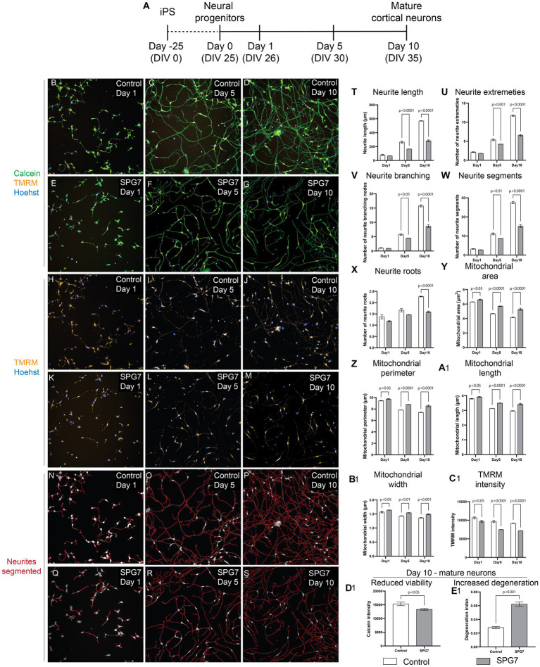
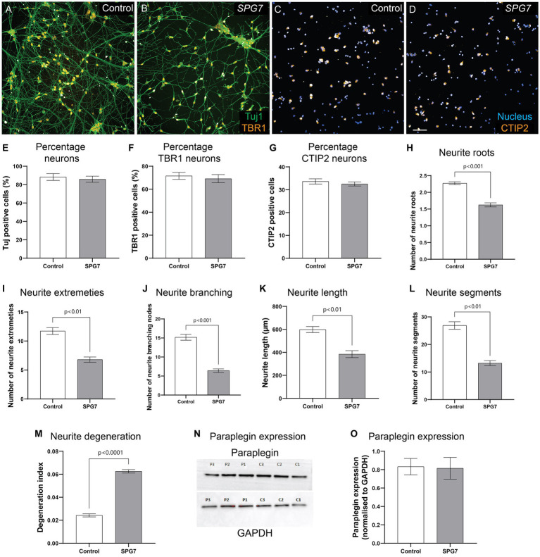
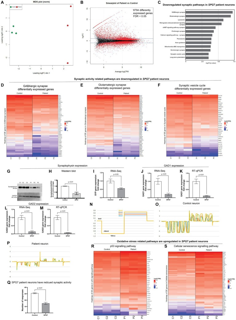
SPG7 is the most common form of autosomal recessive hereditary spastic paraplegia (HSP). There is a lack of HSP-SPG7 human neuronal models to understand the disease mechanism and identify new drug treatments. We generated a human neuronal model of HSP-SPG7 using induced pluripotent stem (iPS) cell technology. We first generated iPS cells from three HSP-SPG7 patients carrying different disease-causing variants and three healthy controls. The iPS cells were differentiated to form neural progenitor cells (NPCs) and then from NPCs to mature cortical neurons. Mitochondrial and neuronal defects were measured using a high throughout imaging and analysis-based assay in live cells. Our results show that compared to control NPCs, patient NPCs had aberrant mitochondrial morphology with increased mitochondrial size and reduced membrane potential. Patient NPCs develop to form mature cortical neurons with amplified mitochondrial morphology and functional defects along with defects in neuron morphology - reduced neurite complexity and length, reduced synaptic gene, protein expression and activity, reduced viability and increased axonal degeneration. Treatment of patient neurons with Bz-423, a mitochondria permeability pore regulator, restored the mitochondrial and neurite morphological defects and mitochondrial membrane potential back to control neuron levels and rescued the low viability and increased degeneration in patient neurons. This study establishes a direct link between mitochondrial and neuronal defects in HSP-SPG7 patient neurons. We present a strategy for testing mitochondrial targeting drugs to rescue neuronal defects in HSP-SPG7 patient neurons.
Keywords: cortical neurons; hereditary spastic paraplegia (HSP); high throughput imaging (HTI); induced pluripotent stem (iPS) cell; mitochondria.
Copyright © 2023 Wali, Li, Liyanage, Kumar, Day and Sue.
Conflict of interest statement
The authors declare that the research was conducted in the absence of any commercial or financial relationships that could be construed as a potential conflict of interest.
Figures

Similar articles
-
Impaired flickering of the permeability transition pore causes SPG7 spastic paraplegia.EBioMedicine. 2020 Nov;61:103050. doi: 10.1016/j.ebiom.2020.103050. Epub 2020 Oct 9. EBioMedicine. 2020. PMID: 33045469 Free PMC article.
-
Mitochondrial Function in Hereditary Spastic Paraplegia: Deficits in SPG7 but Not SPAST Patient-Derived Stem Cells.Front Neurosci. 2020 Aug 20;14:820. doi: 10.3389/fnins.2020.00820. eCollection 2020. Front Neurosci. 2020. PMID: 32973427 Free PMC article.
-
Impaired mitochondrial dynamics underlie axonal defects in hereditary spastic paraplegias.Hum Mol Genet. 2018 Jul 15;27(14):2517-2530. doi: 10.1093/hmg/ddy156. Hum Mol Genet. 2018. PMID: 29726929 Free PMC article.
-
Rescue axonal defects by targeting mitochondrial dynamics in hereditary spastic paraplegias.Neural Regen Res. 2019 Apr;14(4):574-577. doi: 10.4103/1673-5374.248108. Neural Regen Res. 2019. PMID: 30632492 Free PMC article. Review.
-
Pluripotent Stem Cells as a Preclinical Cellular Model for Studying Hereditary Spastic Paraplegias.Int J Mol Sci. 2024 Feb 23;25(5):2615. doi: 10.3390/ijms25052615. Int J Mol Sci. 2024. PMID: 38473862 Free PMC article. Review.
Cited by
-
Neuroaxonal Degeneration as a Converging Mechanism in Motor Neuron Diseases (MNDs): Molecular Insights into RNA Dysregulation and Emerging Therapeutic Targets.Int J Mol Sci. 2025 Aug 7;26(15):7644. doi: 10.3390/ijms26157644. Int J Mol Sci. 2025. PMID: 40806770 Free PMC article. Review.
-
Combining phenomics with transcriptomics reveals cell-type-specific morphological and molecular signatures of the 22q11.2 deletion.Nat Commun. 2025 Jul 9;16(1):6332. doi: 10.1038/s41467-025-61547-x. Nat Commun. 2025. PMID: 40634302 Free PMC article.
-
Single-cell mitochondrial morphomics reveals cellular heterogeneity and predicts complex I, III, and ATP synthase Inhibition responses.Sci Rep. 2025 May 14;15(1):16715. doi: 10.1038/s41598-025-99972-z. Sci Rep. 2025. PMID: 40369116 Free PMC article.
-
Current Applications of Human Pluripotent Stem Cells in Neuroscience Research and Cell Transplantation Therapy for Neurological Disorders.Stem Cell Rev Rep. 2025 May;21(4):964-987. doi: 10.1007/s12015-025-10851-6. Epub 2025 Apr 5. Stem Cell Rev Rep. 2025. PMID: 40186708 Review.
-
Coordinated metabolic responses to cyclophilin D deletion in the developing heart.iScience. 2024 Feb 9;27(3):109157. doi: 10.1016/j.isci.2024.109157. eCollection 2024 Mar 15. iScience. 2024. PMID: 38414851 Free PMC article.
References
LinkOut - more resources
Full Text Sources