

In-cell investigation of the conformational landscape of the GTPase UreG by SDSL-EPR
- PMID: 37766968
- PMCID: PMC10520941
- DOI: 10.1016/j.isci.2023.107855
In-cell investigation of the conformational landscape of the GTPase UreG by SDSL-EPR
Abstract
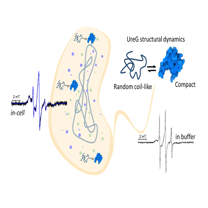
UreG is a cytosolic GTPase involved in the maturation network of urease, an Ni-containing bacterial enzyme. Previous investigations in vitro showed that UreG features a flexible tertiary organization, making this protein the first enzyme discovered to be intrinsically disordered. To determine whether this heterogeneous behavior is maintained in the protein natural environment, UreG structural dynamics was investigated directly in intact bacteria by in-cell EPR. This approach, based on site-directed spin labeling coupled to electron paramagnetic resonance (SDSL-EPR) spectroscopy, enables the study of proteins in their native environment. The results show that UreG maintains heterogeneous structural landscape in-cell, existing in a conformational ensemble of two major conformers, showing either random coil-like or compact properties. These data support the physiological relevance of the intrinsically disordered nature of UreG and indicates a role of protein flexibility for this specific enzyme, possibly related to the regulation of promiscuous protein interactions for metal ion delivery.
Keywords: Analytical chemistry; Microbiology; Spectroscopy.
© 2023 The Authors.
Conflict of interest statement
The authors declare no competing interests.
Figures


Similar articles
-
The conformational response to Zn(II) and Ni(II) binding of Sporosarcina pasteurii UreG, an intrinsically disordered GTPase.J Biol Inorg Chem. 2014 Dec;19(8):1341-54. doi: 10.1007/s00775-014-1191-9. Epub 2014 Sep 9. J Biol Inorg Chem. 2014. PMID: 25200810
-
Nickel and GTP Modulate Helicobacter pylori UreG Structural Flexibility.Biomolecules. 2020 Jul 16;10(7):1062. doi: 10.3390/biom10071062. Biomolecules. 2020. PMID: 32708696 Free PMC article.
-
The relationship between folding and activity in UreG, an intrinsically disordered enzyme.Sci Rep. 2017 Jul 20;7(1):5977. doi: 10.1038/s41598-017-06330-9. Sci Rep. 2017. PMID: 28729736 Free PMC article.
-
Exploring intrinsically disordered proteins using site-directed spin labeling electron paramagnetic resonance spectroscopy.Front Mol Biosci. 2015 May 19;2:21. doi: 10.3389/fmolb.2015.00021. eCollection 2015. Front Mol Biosci. 2015. PMID: 26042221 Free PMC article. Review.
-
Elucidating the design principles of photosynthetic electron-transfer proteins by site-directed spin labeling EPR spectroscopy.Biochim Biophys Acta. 2016 May;1857(5):548-556. doi: 10.1016/j.bbabio.2015.08.009. Epub 2015 Sep 1. Biochim Biophys Acta. 2016. PMID: 26334844 Review.
Cited by
-
Dynamic basis of lipopolysaccharide export by LptB2FGC.Elife. 2024 Oct 7;13:RP99338. doi: 10.7554/eLife.99338. Elife. 2024. PMID: 39374147 Free PMC article.
-
Endogenous Cu(II) Labeling for Distance Measurements on Proteins by EPR.Chemistry. 2024 Dec 23;30(72):e202403160. doi: 10.1002/chem.202403160. Epub 2024 Nov 11. Chemistry. 2024. PMID: 39401409 Free PMC article.
-
Flavoproteins as native and genetically encoded spin probes for in cell ESR spectroscopy.Nat Commun. 2025 Jul 1;16(1):5406. doi: 10.1038/s41467-025-60623-6. Nat Commun. 2025. PMID: 40595551 Free PMC article.
