. 2023 Sep 9;26(10):107863.
doi: 10.1016/j.isci.2023.107863. eCollection 2023 Oct 20.

Stacks of monogalactolipid bilayers can transform into a lattice of water channels

Affiliations

Stacks of monogalactolipid bilayers can transform into a lattice of water channels

Jakub Hryc et al. iScience. .

Abstract

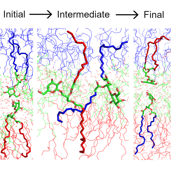
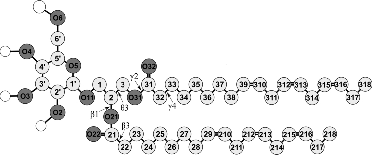
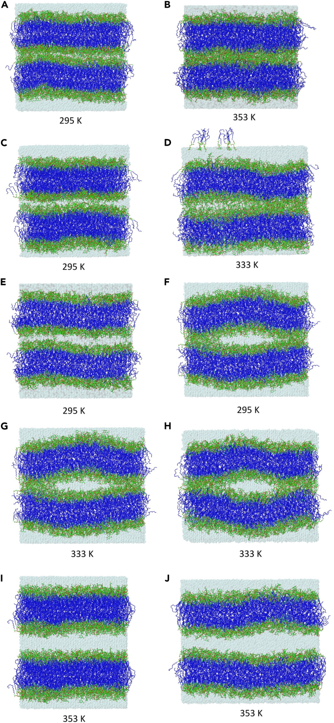
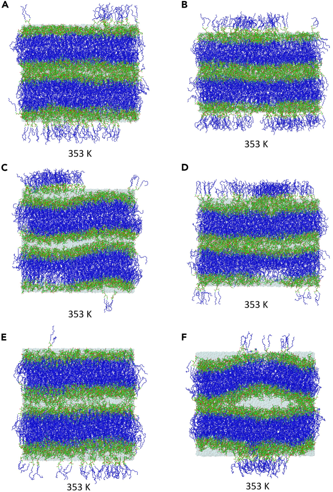
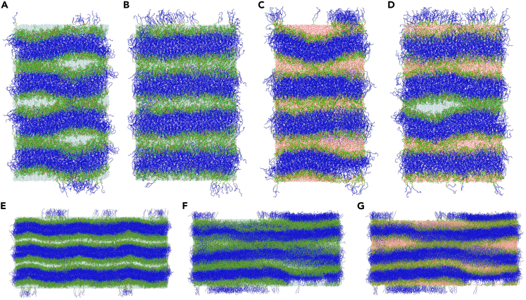
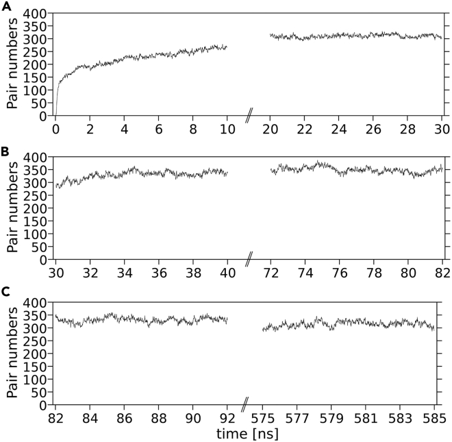
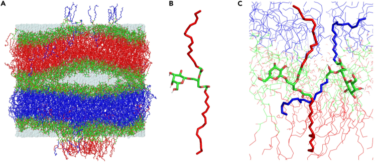
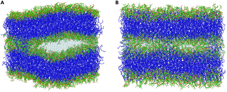
The lipid matrix of thylakoid membranes is a lamellar bilayer, but under a certain condition it can convert locally into a nonlamellar structure. This is possible because one of the main membrane lipids, MGDG, promotes the formation of an inverse hexagonal phase. Here, the spontaneous transformation of aligned hydrated MGDG bilayers into nonlamellar structures is investigated using all-atom molecular dynamics simulation. Previous studies have demonstrated that MGDG polar head groups connect vertically across the interface. In this study, the evolution of the system's initial structure into a lattice of water channels and contacted surfaces created by numerous vertical MGDG connections depended on the width of the hydrating water layers. These widths controlled the bilayers' ability to bend, which was a prerequisite for channel formation. Locally, an intensive exchange of MGDG molecules between apposing bilayer leaflets occurred, although a stable semi-toroidal stalk did not develop.

Keywords: Biophysics; Membrane architecture; Structural biology.


Conflict of interest statement

The authors declare no competing interests.

Figures

None
Graphical abstract
Figure 1
Figure 1
Molecular structures of di-18:3-cis MGDG The acyl chains and glycerol atoms are numbered according to Sundaralingam’s nomenclature (the positions of atoms C1 and C3 are swapped relative to the sn convention). The galactose atoms are numbered according to the IUPAC convention. The galactose carbon atoms are marked with ′ to distinguish them from the C1, C2, and C3 atoms of the glycerol. The chemical symbol for carbon atoms, C, is omitted, and the hydrogen atoms are not shown except for the polar ones shown as empty circles. Oxygen (O) atoms are dark, and the carbon atoms are light gray circles, respectively. Some of the torsion angles discussed in the text are marked, θ3 in the glycerol backbone, β1 and β3 in the β (sn-2) chain, and γ2 and γ4 in the γ (sn-1) chain.
Figure 2
Figure 2
Snapshots of double bilayers with different numbers of water molecules in the inner and the outer water layer (A–J) Initial (A, C, E, G, I) and final, after 585, 700, 320, 820, and 500 ns of simulation (B, D, F, H, J, respectively) structures of double bilayers. W8-30 (A, B), W12-30 (C, D), W15-30 (295 K, E, F), W15-30 (333 K, G, H), and W30-30 (I, J) (see text). The atoms are represented in standard colors, except for the acyl chain carbon atoms, which are dark blue. The water is shown as a transparent blue surface. The MGDG hydrogen atoms are not shown. The temperature corresponding to each structure is given below the structure.
Figure 3
Figure 3
Snapshots of double bilayers with the same number of water molecules in the inner and the outer water layer (A–F) Initial (A, C, E) and final, after 1000, 500, 1000 ns of simulation (B, D, F, respectively) structures of double bilayers. W8-8 (A, B), W12-12 (C, D), W15-15 (E, F). Other details as in Figure 2.
Figure 4
Figure 4
Snapshots of quadruple and triple bilayers (A–G) Initial (A, B, E) and final, after 1000 ns of simulation (C, D, F, G) structures of 2xW15-15 (A, B, C, D) and 6xW15-15-15 (E, F, G) systems. To show the direction of the water channels in 2xW15-15, (A, C) are the front (x,z-plane) and (B, D) are the side (y,z-plane) views. (E, F, G) are the front views of 6xW15-15-15. In (A, B, E, F, and partially in D) the water is shown as a transparent blue surface; to show the water-filled channels better, in (C, G, and partially in D) the water molecules are shown explicitly to expose the water-filled caves better. Other details as in Figure 2.
Figure 5
Figure 5
Time profiles of the number of vertical MGDG pairs linking the inner leaflets of W8-30 (A–C) (A) 295 K (30-ns simulation, aniso); (B) 333 K (52.5-ns simulation, aniso); (C) 353 K (500-ns simulation, semi). At each temperature, the pairs were counted during the first and the last 10-ns period of simulation every 1 ps; each point on the plot is a 10-ps average. Each bilayer leaflet contained 225 MGDG molecules. See also Figures S3–S7 and Table 3.
Figure 6
Figure 6
Exchanging of MGDG molecules between leaflets (A) Snapshot of W15-15 at the end of 1000-ns simulation. Acyl chains are colored red for the upper bilayer and blue for the lower bilayer to make the exchanging MGDG molecules more visible. (B) An MGDG molecule from the “red” bilayer in the splayed-chain (extended) conformation—it remains in this conformation for 48 ns. (C) Two simultaneously exchanging MGDG molecules, red and blue; each is in the extended conformation with chains embedded in the opposing leaflet and head groups in the interface. See also Figures S8 and S9.
Figure 7
Figure 7
Local exchange of MGDG molecules across the interface (A and B) The (A) front and (B) side view of the outer leaflets of 2xW15-15. Both in (A) and (B) acyl chains of several MGDG molecules can be seen in the interfacial region. Other details as in Figure 2. See also Figure S8.
Figure 8
Figure 8
Hydration of a MGDG molecule during swapping RDFs of water molecules relative to the MGDG β-chain (blue) and γ-chain (green) carbon atoms and head group oxygen atoms (red) of an MGDG molecule during swapping between leaflets.

Similar articles

References

    1. Dörmann P., Benning C. Galactolipids rule in seed plants. Trends Plant Sci. 2002;7:112–118. doi: 10.1016/s1360-1385(01)02216-6. - DOI - PubMed
    1. Kobayashi K. Role of membrane glycerolipids in photosynthesis, thylakoid biogenesis and chloroplast development. J. Plant Res. 2016;129:565–580. doi: 10.1007/s10265-016-0827-y. - DOI - PMC - PubMed
    1. Ohlrogge J., Browse J. Lipid biosynthesis. Plant Cell. 1995;7:957–970. doi: 10.1105/tpc.7.7.957. - DOI - PMC - PubMed
    1. Lee A.G. Membrane lipids: it's only a phase. Curr. Biol. 2000;10:R377–R380. doi: 10.1016/s0960-9822(00)00477-2. - DOI - PubMed
    1. Shipley G.G., Green J.P., Nichols B.W. The phase behavior of monogalactosyl, digalactosyl, and sulphoquinovosyl diglycerides. Biochim. Biophys. Acta. 1973;311:531–544. doi: 10.1016/0005-2736(73)90128-4. - DOI - PubMed

LinkOut - more resources