

Age, Gender, Body Mass Index, and Foot Loading During Gait
- PMID: 37767006
- PMCID: PMC10521294
- DOI: 10.1177/24730114231198524
Age, Gender, Body Mass Index, and Foot Loading During Gait
Abstract
Background: The aim was to analyze changes in normal functional parameters of gait analysis by aging, sex, and body mass index (BMI).
Methods: A cross-sectional study with a consecutive sample of asymptomatic subjects was performed between 2014 and 2020. Primary outcomes were time and force parameters (contact time and center of force [CoF] time), in the heel, midfoot, and metatarsal areas, measured using an in-office force platform.
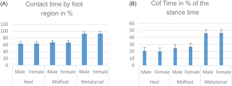
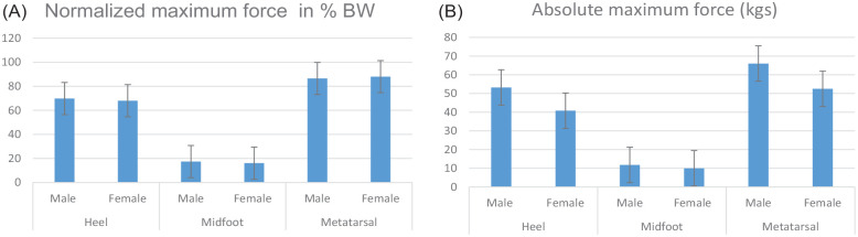
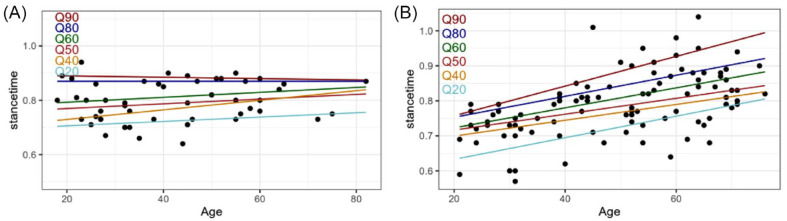
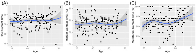
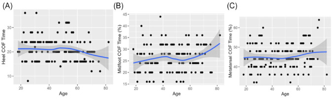
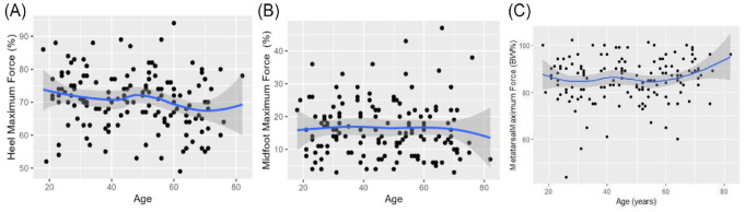
Results: A total of 156 subjects (312 feet) were included, including 67% of women with a mean age of 47 years. The mean of total contact time was similar in males and females (P = .695) and across BMI (P = .413). Contact time did not show differences by region (P = .648 heel, P = .286 midfoot, and P = .690 metatarsal). CoF time in the heel and metatarsal areas did not change between males and females (P = .288 and P = .879, respectively); meanwhile, it was different in midfoot (P = .002). Maximum force showed a reduction between sexes in the heel (P = .039) but did not in the midfoot and metatarsal areas. By age, differences were detected in the heel and metatarsal areas in females (P = .002 and P = .001) and the metatarsal area in males (P = .001). According to the age groups, total contact time increased in females (P = .001) but not in males (P = .018), and no differences were detected between foot areas. In females, CoF time did not change either foot areas or age groups. In males, CoF time values increased in the midfoot area in the older group (P = .001).
Conclusion: Time variables did not change by foot region, independent of age, sex, and BMI. Heel maximum force decreased in females, probably linked to adaptive phenomena by aging. The midfoot remains stable, and acts as an undamaged "bridge." These parameters could be interpreted as normal in asymptomatic subjects.
Level of evidence: Level III, diagnostic and prognostic.
Keywords: asymptomatic patients; center of force; contact time; foot functional parameters; force platform; gait analysis; ground reaction force; maximum force.
© The Author(s) 2023.
Conflict of interest statement
The author(s) declared no potential conflicts of interest with respect to the research, authorship, and/or publication of this article. ICMJE forms for all authors are available online.
Figures
References
-
- Anderson FC, Pandy MG. Individual muscle contributions to support in normal walking. Gait Posture. 2003;17(2):159-169. - PubMed
-
- Castellini JL. Parametros biomecanicos de la funcion del pie medidos en el consultorio del especialista en Ortopedia y Traumatologia [Biomechanical parameters of foot function measured in an orthopedic and traumatology specialist’s office]. Rev Asoc Argent Ortop Argent. 2022;87(6):756-764.
LinkOut - more resources
Full Text Sources
Research Materials