

Accuracy of radiographic measurements of fracture-induced deformity in the distal radius
- PMID: 37767057
- PMCID: PMC10521277
- DOI: 10.1177/20584601231205986
Accuracy of radiographic measurements of fracture-induced deformity in the distal radius
Abstract
Background: Management of the distal radius fracture (DRF) is to some extent based on radiographic characterization of fracture displacement. It remains unclear, however, if the measurements used to quantify displacement are accurate.
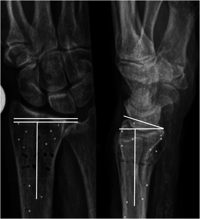
Purpose: To quantify accuracy of two radiographic measurements: dorsal/volar tilt and fracture compression, measured indirectly as ulnar variance (UV), using radiostereometric analyses (RSA) as reference standard.
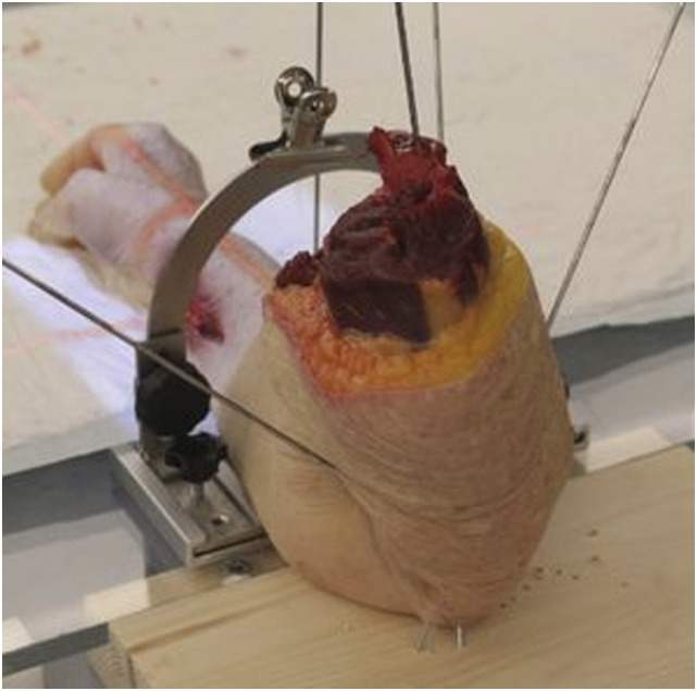
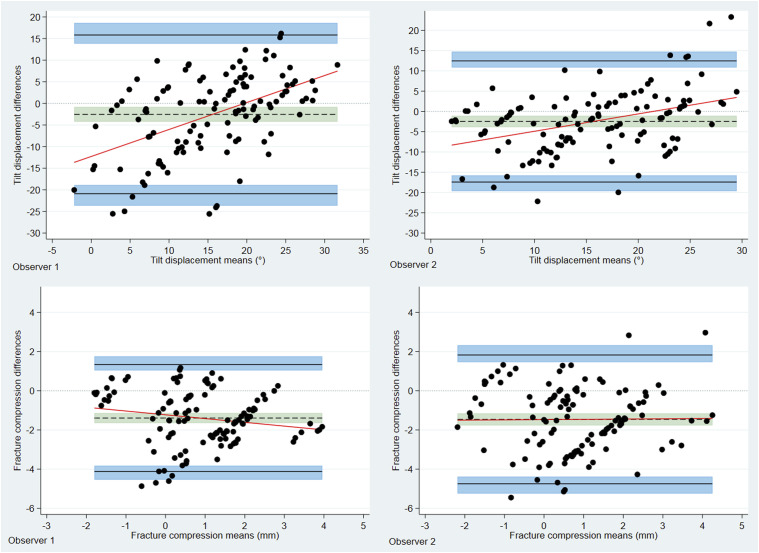
Material and methods: Twenty-one fresh frozen non-fractured human cadaveric forearms (right = 11, left = 10) were thawed and eligible for inclusion. The forearms were mounted on a custom made platform that allowed for controlled forearm rotation, and they underwent two rounds of imaging (both rounds consisted of RSA and radiographs). In round one, the non-fractured forearms were radiographed. In round two, artificial DRF´s with compression and dorsal angulation were created and imaging procedures repeated. Change in tilt and UV between the non-fractured and later fractured forearms was defined as fracture-induced deformity. Deformity was measured radiographically and additionally calculated using RSA. Bland Altman analyses were used to estimate agreement between radiographically measured, and RSA calculated, fracture-induced deformity.
Results: Our results indicated that radiographs underestimate the amount of fracture-induced deformity. Mean measured differences (bias) in dorsal tilt deformity between radiographs and RSA were -2.5° for both observers. The corresponding values for UV were -1.4 mm and -1.5 mm.
Conclusion: Quantifying fracture-induced deformity on radiographs underestimated the actual deformity when compared to RSA calculated deformity. These findings suggest that clinicians, at least in part, base fracture management and potentially corrective surgery on inaccurate measurements.
Keywords: distal radius fracture; dorsal tilt; measurements accuracy; radial inclination; ulnar variance.
© The Author(s) 2023.
Conflict of interest statement
The author(s) declared no potential conflicts of interest with respect to the research, authorship, and/or publication of this article.
Figures
References
-
- Danish Department of Health . National clinical guideline on the treatment of distal radial fractures. Available at: https://www.sst.dk/-/media/Udgivelser/2014/NKR-H%C3%A5ndledsn%C3%A6re-un... (2022, accessed 25 March 2022).
-
- Treatment of distal radius fractures in adults - Norwegian orthopaedic association - The Norwegian medical association. Available at: https://files.magicapp.org/guideline/ac10868f-c18b-462d-978d-cb53a5959fd... (2023, accessed 6 May 2023).
-
- Katz MA, Beredjiklian PK, Bozentka DJ, et al. Computed tomography scanning of intra-articular distal radius fractures: does it influence treatment? J Hand Surg AM 2001; 26: 415–421. - PubMed
-
- Värttinäluun alaosan murtuma (rannemurtuma) . Available at: https://www.kaypahoito.fi/hoi50109 (2022, Accessed 25 March 2022).
LinkOut - more resources
Full Text Sources
