

Establishment of a uniform histological evaluation method for early stage osteophytes in the destabilization of the medial meniscus mouse model
- PMID: 37767107
- PMCID: PMC10520448
- DOI: 10.1016/j.ocarto.2023.100409
Establishment of a uniform histological evaluation method for early stage osteophytes in the destabilization of the medial meniscus mouse model
Abstract
Background: Osteophyte formation is attracting attention as an early-stage pathology of knee osteoarthritis (OA). Although osteophyte formation is understood as a defense response to joint instability, its role and impact on OA remain largely unknown. Many studies have been conducted using the surgical destabilization of the medial meniscus (DMM) mouse model, but there are few standard evaluation methods, especially in the histological evaluation of early-stage osteophytes. The purpose of this study was to establish a reproducible and uniform method for histological evaluation of characteristics of early osteophyte formation in the DMM mouse model.
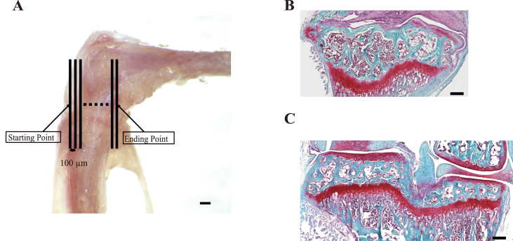
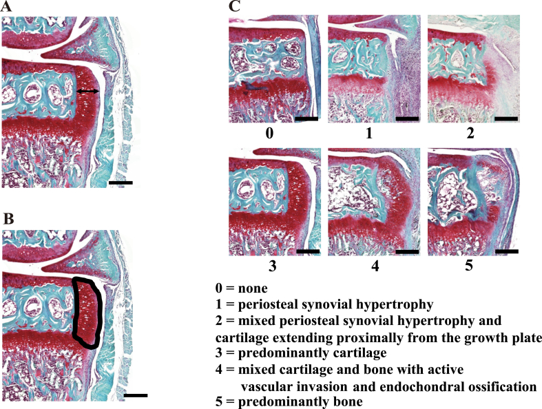
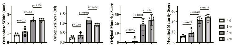
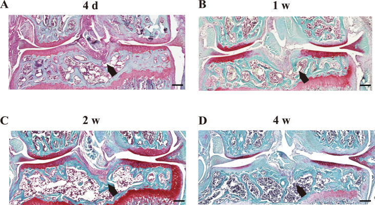
Methods: Male mice were operated with DMM at 12 weeks old and histologically evaluated at 4 days and 1, 2 and 4 weeks after DMM. Osteophyte Width, Osteophyte Area, and Original and Modified Maturity Scores were used to evaluate osteophytes for all sections.
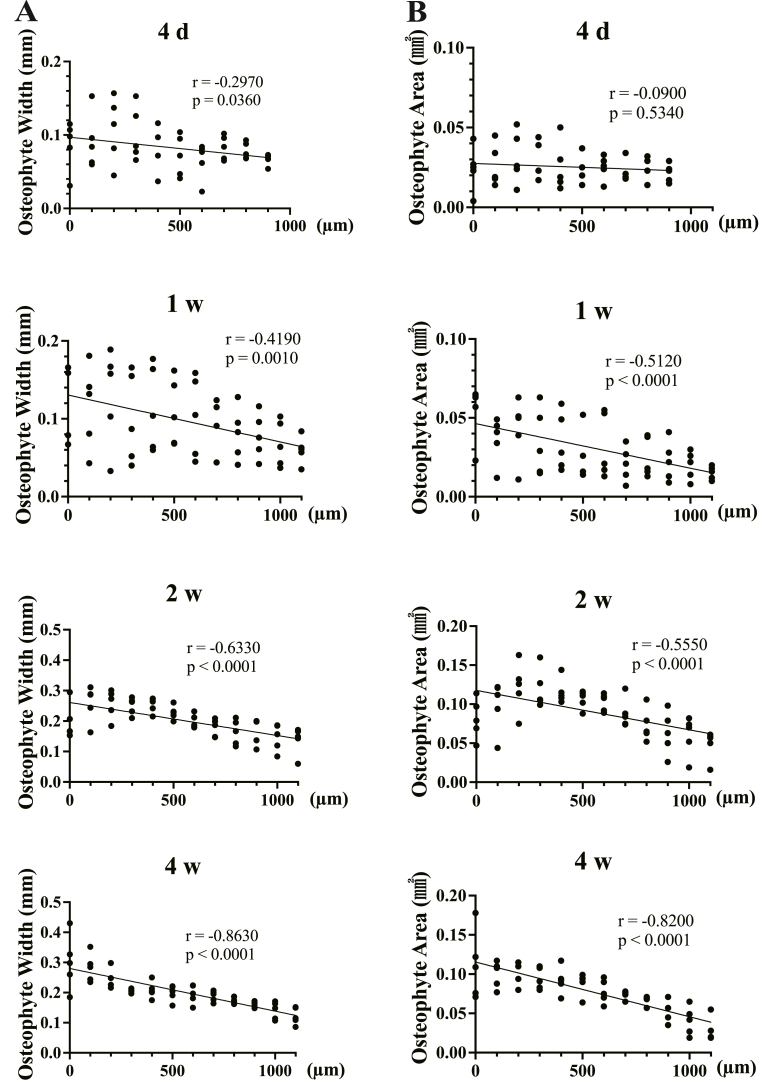
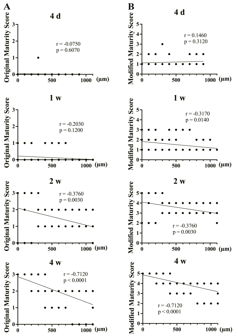
Results: Osteophyte Width, Osteophyte Area and Maturity Scores were all greater anteriorly than posteriorly in the knee joint. The Modified Maturity Score was more strongly correlated with position than the Original Maturity Score, and could be used to evaluate early-stage osteophyte formation.
Conclusion: The Modified Maturity Score as well as Osteophyte Width and Area at the section of the anterior cruciate ligament (ACL) attachment site can provide a reproducible evaluation method to histologically assess the early-stage osteophyte formation in the DMM mouse model.
Keywords: DMM mouse model; Osteoarthritis; Osteophyte; Osteophyte maturity score; Synovium.
© 2023 The Author(s).
Conflict of interest statement
All authors declare that they have no conflict of interest.
Figures
Similar articles
-
Association of medial meniscal extrusion with medial tibial osteophyte distance detected by T2 mapping MRI in patients with early-stage knee osteoarthritis.Arthritis Res Ther. 2017 Sep 12;19(1):201. doi: 10.1186/s13075-017-1411-0. Arthritis Res Ther. 2017. PMID: 28899407 Free PMC article.
-
Associations between biomarkers and histological assessment in individual animals in a destabilization of the medial meniscus (DMM) model of osteoarthritis (OA).Acta Orthop Belg. 2021 Dec;87(4):713-721. doi: 10.52628/87.4.16. Acta Orthop Belg. 2021. PMID: 35172438
-
Relationship between histological changes of the anterior cruciate ligament and knee function in osteoarthritis patients.Orthop Traumatol Surg Res. 2022 Dec;108(8):103341. doi: 10.1016/j.otsr.2022.103341. Epub 2022 May 25. Orthop Traumatol Surg Res. 2022. PMID: 35643361
-
Reduction of knee joint load suppresses cartilage degeneration, osteophyte formation, and synovitis in early-stage osteoarthritis using a post-traumatic rat model.PLoS One. 2021 Jul 16;16(7):e0254383. doi: 10.1371/journal.pone.0254383. eCollection 2021. PLoS One. 2021. PMID: 34270585 Free PMC article.
-
Differential patterns of pathology in and interaction between joint tissues in long-term osteoarthritis with different initiating causes: phenotype matters.Osteoarthritis Cartilage. 2020 Jul;28(7):953-965. doi: 10.1016/j.joca.2020.04.009. Epub 2020 Apr 30. Osteoarthritis Cartilage. 2020. PMID: 32360537
Cited by
-
In Vivo and In Vitro Evaluation of the Feasibility and Safety Profiles of Intraarticular Transplantation of Mitochondria for Future Use as a Therapy for Osteoarthritis.Cells. 2025 Jan 21;14(3):151. doi: 10.3390/cells14030151. Cells. 2025. PMID: 39936943 Free PMC article.
-
Assessment of Feasibility of the M2 Macrophage-Based Adoptive Gene Transfer Strategy for Osteoarthritis with a Mouse Model.Cells. 2025 Jul 11;14(14):1067. doi: 10.3390/cells14141067. Cells. 2025. PMID: 40710320 Free PMC article.
References
-
- Chiang H., Jiang C.C. Repair of articular cartilage defects: review and perspectives. J. Formos. Med. Assoc. 2009;108:87–101. - PubMed
-
- Hunter D.J., Bierma-Zeinstra S. Osteoarthritis. Lancet. 2019;393:1745–1759. - PubMed
-
- Adams J.G., McAlindon T., Dimasi M., Carey J., Eustace S. Contribution of meniscal extrusion and cartilage loss to joint space narrowing in osteoarthritis. Clin. Radiol. 1999;54:502–506. - PubMed
-
- Berthiaume M.J., Raynauld J.P., Martel-Pelletier J., Labonté F., Beaudoin G., Bloch D.A., et al. Meniscal tear and extrusion are strongly associated with progression of symptomatic knee osteoarthritis as assessed by quantitative magnetic resonance imaging. Ann. Rheum. Dis. 2005;64:556–563. - PMC - PubMed
LinkOut - more resources
Full Text Sources
