

Mature teratoma of conus medullaris: A case report and review of literature
- PMID: 37767146
- PMCID: PMC10520346
- DOI: 10.1002/ccr3.7966
Mature teratoma of conus medullaris: A case report and review of literature
Abstract
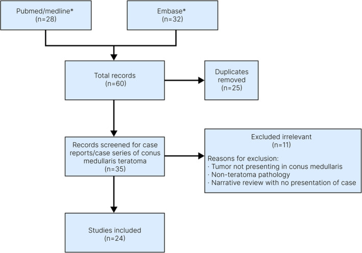
In conus medullaris, mature teratomas are rare. We report a case of a 40-year-old man who presented with urinary incontinence, low back pain, and muscle weakness. Magnetic resonance imaging revealed a mass in conus medullaris (T11-L1), further confirmed as a mature teratoma by pathological examination. We identified 63 cases of conus medullaris teratoma over the past two decades by systematically analyzing the case reports. Findings demonstrated that most cases were diagnosed in the fourth decade of life, with the majority of cases (57.6%) being male. Lower back pain, radiating pain in the extremities, hypoesthesia, and urinary dysfunction are the most common clinical presentations among patients with teratoma of conus medullaris. Mature teratoma is the dominant pathologic subtype of teratomas in this region, comprising more than 95% of cases. Our case highlights the importance of considering spinal teratoma as a differential diagnosis in patients presenting with urinary incontinence and lumbar pain.
Keywords: conus medullaris; mature teratoma; spinal tumor.
© 2023 The Authors. Clinical Case Reports published by John Wiley & Sons Ltd.
Conflict of interest statement
The authors declare that no competing and financial interests exist.
Figures


Similar articles
-
Exophytic intramedullary mature teratoma of the conus medullaris: case report and review of the literature.Cent Eur Neurosurg. 2009 Aug;70(3):154-60. doi: 10.1055/s-0028-1082062. Epub 2009 Aug 21. Cent Eur Neurosurg. 2009. PMID: 19701875
-
Coexistence of spinal teratoma of the conus medullaris and arteriovenous malformation in an adult: a case report.Turk Neurosurg. 2012;22(4):510-4. doi: 10.5137/1019-5149.JTN.3961-10.1. Turk Neurosurg. 2012. PMID: 22843478
-
Conus Medullaris Teratoma with Utilization of Fiber Tractography: Case Report.J Neurol Surg Rep. 2015 Jul;76(1):e183-7. doi: 10.1055/s-0035-1555134. Epub 2015 Jun 12. J Neurol Surg Rep. 2015. PMID: 26251802 Free PMC article.
-
Cystic mature teratoma of the filum terminale in an adult. Case report and review of the literature.Neurocirugia (Astur). 2004 Jun;15(3):290-3. doi: 10.1016/s1130-1473(04)70486-8. Neurocirugia (Astur). 2004. PMID: 15239016 Review.
-
Adult intradural extramedullary teratoma of the spinal cord: A case presentation.Clin Neurol Neurosurg. 2018 Dec;175:54-56. doi: 10.1016/j.clineuro.2018.10.010. Epub 2018 Oct 16. Clin Neurol Neurosurg. 2018. PMID: 30384116 Review.
Cited by
-
Conus Medullaris Teratoma in an Adolescent: A Case Report and Literature Review.Cureus. 2025 Jul 25;17(7):e88758. doi: 10.7759/cureus.88758. eCollection 2025 Jul. Cureus. 2025. PMID: 40861551 Free PMC article.
References
-
- Agrawal M, Uppin MS, Patibandla MR, et al. Teratomas in central nervous system: a clinico‐morphological study with review of literature. Neurol India. 2010;58(6):841‐846. - PubMed
-
- Schmidt RF, Casey JP, Gandhe AR, Curtis MT, Heller JE. Teratoma of the spinal cord in an adult: report of a rare case and review of the literature. J Clin Neurosci. 2017;36:59‐63. - PubMed
-
- Lim EA, Alves CA, Picariello S, et al. Neuroimaging of paediatric pineal, sellar and suprasellar tumours: a guide to differential diagnosis. Childs Nerv Syst. 2022;38:33‐50. - PubMed
-
- Liu Z, Lv X, Wang W, et al. Imaging characteristics of primary intracranial teratoma. Acta Radiol. 2014;55(7):874‐881. - PubMed
Publication types
LinkOut - more resources
Full Text Sources
Miscellaneous
