

Super Enhancer Regulatory Gene FYB1 Promotes the Progression of T Cell Acute Lymphoblastic Leukemia by Activating IGLL1
- PMID: 37767202
- PMCID: PMC10522422
- DOI: 10.1155/2023/3804605
Super Enhancer Regulatory Gene FYB1 Promotes the Progression of T Cell Acute Lymphoblastic Leukemia by Activating IGLL1
Abstract
Background: Arising from T progenitor cells, T-cell acute lymphoblastic leukemia (T-ALL) is an aggressive hematologic malignant tumor, accounting for 15% of childhood ALL and 25% of adult ALL. Composing of putative enhancers in close genomic proximity, super enhancer (SE) is critical for cell identity and the pathogenesis of multiple cancers. Belonging to the cytosolute linker protein group, FYB1 is essential for TCR signaling and extensively studied in terms of tumor pathogenesis and metastasis. Dissecting the role of FYN binding protein 1 (FYB1) in T-ALL holds the potential to improve the treatment outcome and prognosis of T-ALL.
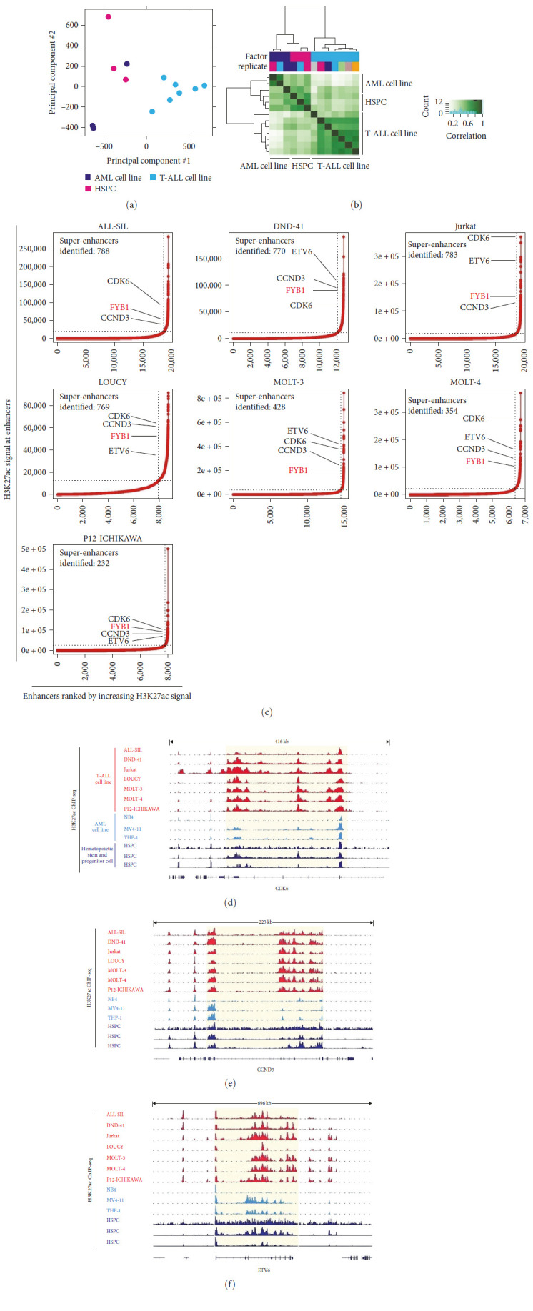
Methods: In this study, SEs were explored using public H3K27ac ChIP-seq data derived from T-ALL cell lines, AML cell lines and hematopoietic stem and progenitor cells (HSPCs). Downstream target of FYB1 gene was identified by RNA-seq. Effects of shRNA-mediated downregulation of FYB1 and immunoglobulin lambda-like polypeptide 1 (IGLL1) on self-renewal of T-ALL cells were evaluated in vitro and/or in vivo.
Results: As an SE-driven gene, overexpression of FYB1 was observed in T-ALL, according to the Cancer Cell Line Encyclopedia database. In vitro, knocking down FYB1 led to comprised growth and enhanced apoptosis of T-ALL cells. In vivo, downregulation of FYB1 significantly decreased the disease burden by suppressing tumor growth and improved survival rate. Knocking down FYB1 resulted in significantly decreased expression of IGLL1 that was also an SE-driven gene in T-ALL. As a downstream target of FYB1, IGLL1 exerted similar role as FYB1 in inhibiting growth of T-ALL cells.
Conclusion: Our results suggested that FYB1 gene played important role in regulating self-renewal of T-ALL cells by activating IGLL1, representing a promising therapeutic target for T-ALL patients.
Copyright © 2023 Kunlong Zhang et al.
Conflict of interest statement
The authors declare that they have no conflicts of interest.
Figures


Similar articles
-
FYB1-targeted modulation of CAPG promotes AML progression.Mol Cell Biochem. 2025 Feb;480(2):985-999. doi: 10.1007/s11010-024-04992-4. Epub 2024 May 3. Mol Cell Biochem. 2025. PMID: 38700746 Free PMC article.
-
Super-enhancer profiling identifies novel critical and targetable cancer survival gene LYL1 in pediatric acute myeloid leukemia.J Exp Clin Cancer Res. 2022 Jul 16;41(1):225. doi: 10.1186/s13046-022-02428-9. J Exp Clin Cancer Res. 2022. PMID: 35842703 Free PMC article.
-
[Expression of IGLL1 Gene and Its Clinical Significance in Pediatric T-ALL].Zhongguo Shi Yan Xue Ye Xue Za Zhi. 2023 Aug;31(4):999-1004. doi: 10.19746/j.cnki.issn.1009-2137.2023.04.011. Zhongguo Shi Yan Xue Ye Xue Za Zhi. 2023. PMID: 37551468 Chinese.
-
[Understanding of molecular pathogenesis of T-cell leukemia by super-enhancer profiling].Rinsho Ketsueki. 2018;59(7):899-908. doi: 10.11406/rinketsu.59.899. Rinsho Ketsueki. 2018. PMID: 30078801 Review. Japanese.
-
Super enhancers as master gene regulators in the pathogenesis of hematologic malignancies.Biochim Biophys Acta Rev Cancer. 2022 Mar;1877(2):188697. doi: 10.1016/j.bbcan.2022.188697. Epub 2022 Feb 9. Biochim Biophys Acta Rev Cancer. 2022. PMID: 35150791 Review.
Cited by
-
Core transcriptional regulatory circuitry molecule ZNF217 promotes AML cell proliferation by up-regulating MYB.Int J Biol Sci. 2025 Feb 18;21(5):1966-1983. doi: 10.7150/ijbs.103211. eCollection 2025. Int J Biol Sci. 2025. PMID: 40083704 Free PMC article.
-
Analysis of genomic selection characteristics of local cattle breeds in Gansu.BMC Genomics. 2025 Jul 1;26(1):574. doi: 10.1186/s12864-025-11753-0. BMC Genomics. 2025. PMID: 40597588 Free PMC article.
-
FYB1-targeted modulation of CAPG promotes AML progression.Mol Cell Biochem. 2025 Feb;480(2):985-999. doi: 10.1007/s11010-024-04992-4. Epub 2024 May 3. Mol Cell Biochem. 2025. PMID: 38700746 Free PMC article.
-
Deep hematologic response to RD treatment in patients with multiple myeloma is associated with overexpression of IL-17R in CD138+ plasma cells.Sci Rep. 2024 Oct 9;14(1):23559. doi: 10.1038/s41598-024-74558-3. Sci Rep. 2024. PMID: 39384864 Free PMC article.
References
LinkOut - more resources
Full Text Sources
Research Materials
Miscellaneous
