

Solitary Intestinal Lymphangiectasia Causing Transient Intussusception
- PMID: 37767268
- PMCID: PMC10521876
- DOI: 10.7759/cureus.44206
Solitary Intestinal Lymphangiectasia Causing Transient Intussusception
Abstract
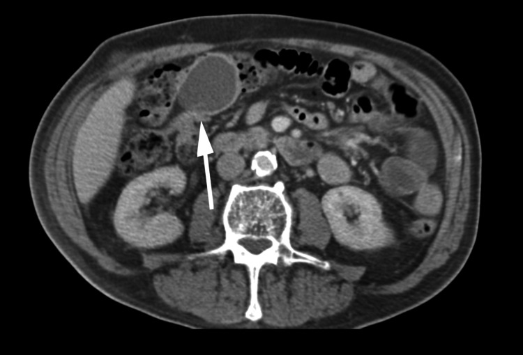
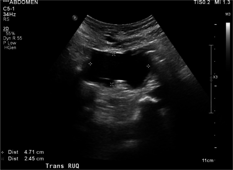
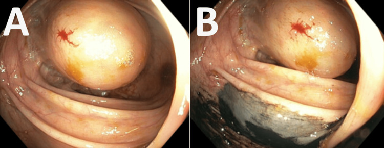
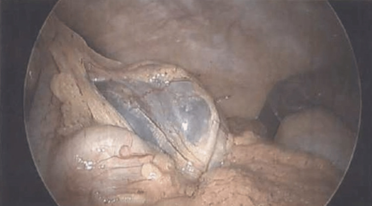
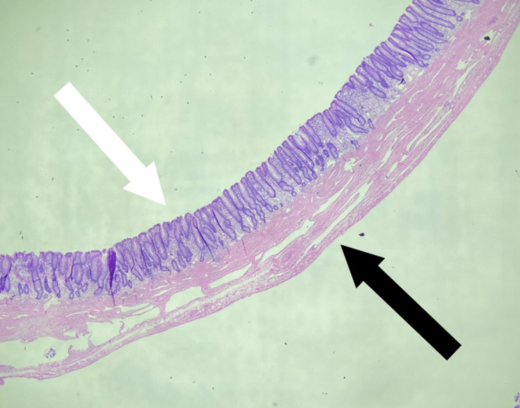
Lymphangiectasia is the benign malformation of lymphatic channels associated with either focal or diffuse dilation of vessels and impaired lymph drainage. This malformation has the potential to create a cystic mass due to the accumulation of lymphatic fluid. While rare in adults, intussusception, the telescoping of the proximal bowel into the distal bowel, can be caused by a mass within the bowel. In this case, a near-obstructing cystic colon mass developed in a 74-year-old man; this was later found to be a large lymphangiectasia. In addition, this near-obstructing colonic lymphangiectasia served as the lead point in a colo-colonic intussusception. Due to this complication, the mass was immediately removed by a laparoscopic oncologic right-extended hemicolectomy which proved to be both diagnostic and therapeutic.
Keywords: bowel obstruction; intussusception; lymph; lymphangiectasia; lymphatic malformation; lymphatics; colonoscopy.
Copyright © 2023, Persin et al.
Conflict of interest statement
The authors have declared that no competing interests exist.
Figures

Similar articles
-
Adult colo-colonic intussusception caused by congenital bands: A case report and literature review.Int J Surg Case Rep. 2016;26:88-92. doi: 10.1016/j.ijscr.2016.07.019. Epub 2016 Jul 22. Int J Surg Case Rep. 2016. PMID: 27475114 Free PMC article.
-
Giant pedunculated colonic lipoma causing colo-colic intussusception in a patient with mechanical ileus.Ann Ital Chir. 2020 Mar 3;9:S2239253X20032296. Ann Ital Chir. 2020. PMID: 32989208
-
Colo-colonic intussusception as a rare complication of colonoscopy with polypectomy: Two case reports.World J Gastrointest Surg. 2024 Jun 27;16(6):1939-1947. doi: 10.4240/wjgs.v16.i6.1939. World J Gastrointest Surg. 2024. PMID: 38983333 Free PMC article.
-
Ileal polypoid lymphangiectasia bleeding diagnosed and treated by double balloon enteroscopy.World J Gastroenterol. 2013 Dec 7;19(45):8440-4. doi: 10.3748/wjg.v19.i45.8440. World J Gastroenterol. 2013. PMID: 24363538 Free PMC article. Review.
-
Cystic colon duplication causing intussusception in a 25-year-old man: report of a case and review of the literature.BMC Surg. 2010 Jun 23;10:19. doi: 10.1186/1471-2482-10-19. BMC Surg. 2010. PMID: 20573256 Free PMC article. Review.
References
-
- The diagnosis and management of adult intussusception. Begos DG, Sandor A, Modlin IM. https://doi.org/10.1016/S0002-9610. Am J Surg. 1997;173:88–94. - PubMed
-
- Primary intestinal lymphangiectasia: minireview. Ingle SB, Hinge Ingle CR. https://doi.org/10.12998/wjcc.v2.i10.528. World J Clin Cases. 2014;2:528–533. - PMC - PubMed
-
- The role of the gastrointestinal system in "idiopathic hypoproteinemia". Waldmann TA, Steinfeld JL, Dutcher TF, Davidson JD, Gordon RS. Gastroenterology. 41:197–207. - PubMed
-
- Primary intestinal lymphangiectasia (Waldmann's disease) Vignes S, Bellanger J. https://doi.org/10.1186/1750-1172-3-5. Orphanet J Rare Dis. 2008;3:5. - PMC - PubMed
-
- Generalized lymphangiomatosis. Marom EM, Moran CA, Munden RF. https://doi.org/10.2214/ajr.182.4.1821068. AJR Am J Roentgenol. 2004;182:1068. - PubMed
Publication types
LinkOut - more resources
Full Text Sources