

Three-dimensional morphometric analysis of cranial sutures - A novel approach to quantitative analysis
- PMID: 37767331
- PMCID: PMC10520544
- DOI: 10.1016/j.bonr.2023.101714
Three-dimensional morphometric analysis of cranial sutures - A novel approach to quantitative analysis
Abstract
Objective: Differences in complexity of cranial suture forms on the endocranial (i.e., deep) and ectocranial (i.e., superficial) skull surfaces have been noted in the literature, indicating through thickness three-dimensional (3D) suture variability depending on the chosen section and necessity for considering the complete 3D structure in many cases. This study aims to evaluate the variability of suture morphology through the skull thickness using a rat model, and to provide more robust metrics and methodologies to analyze suture morphology.
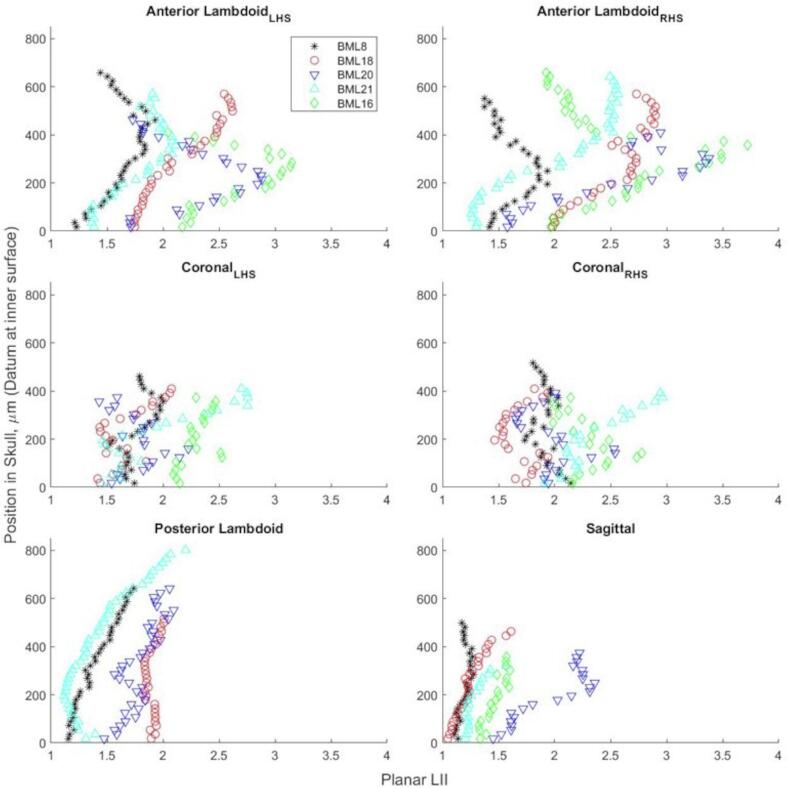
Design: X-ray micro-computed tomographic (μCT) imaging methods were utilized in order to provide internal structure information. Methods were developed to isolate and analyze sutures widths and linear interdigitation index (LII) values on each adjacent offset transverse plane of the μCT datasets. LII was defined as the curved path length of the suture divided by the linear length between the ends of the region of interest. Scans were obtained on 15 female rats at ages of 16, 20, and 24 weeks (n = 5/age). Samples were imaged at 18 μm resolutions with 90 kV source voltage, 278 μA source amperage, and 0.7° increments. Suture widths and LII values were compared using a Kruskal-Wallis test.
Results: 3D variability in local suture widths within individuals, as well as through thickness variabilities in planar widths and LII was observed. Kruskal-Wallis tests for bulk through thickness averaged suture widths and LII were found to be statistically insignificant, despite clear geometric differences through suture thicknesses.
Conclusion: Although the bulk morphometric variability between age groups was found to be statistically insignificant, the 3D variability within individuals point to the importance of analyzing suture form using 3D metrics when studying suture development, response to functional activity, or morphometry in general.
Keywords: Computed tomography; Cranial suture; Image analysis; Linear interdigitation index; Morphology.
© 2023 The Authors.
Conflict of interest statement
The authors declare that they have no known competing financial interests or personal relationships that could have appeared to influence the work reported in this paper.
Figures


References
-
- Bishara S.E., Staley R.N. Maxillary expansion: clinical implications. Am. J. Orthod. Dentofacial Orthop. 1987;91:3–14. - PubMed
-
- Byron C.D. Role of the osteoclast in cranial suture waveform patterning. Anatatomical Record - Part A. Discov. Mol. Cell. Evol. Biol. 2006;288:552–563. - PubMed
-
- Byron C.D. Cranial suture morphology and its relationship to diet in cebus. J. Hum. Evol. 2009;57:649–655. - PubMed
-
- Cohen M.M. Sutural biology and the correlates of craniosynostosis. Am. J. Med. Genet. 1993;47:581–616. - PubMed
LinkOut - more resources
Full Text Sources
