. 2023 Oct 1;150(19):dev201633.
doi: 10.1242/dev.201633. Epub 2023 Oct 2.

Chemokine-like Orion is involved in the transformation of glial cells into phagocytes in different developmental neuronal remodeling paradigms

Affiliations

Chemokine-like Orion is involved in the transformation of glial cells into phagocytes in different developmental neuronal remodeling paradigms

Clarisse Perron et al. Development. .

Abstract

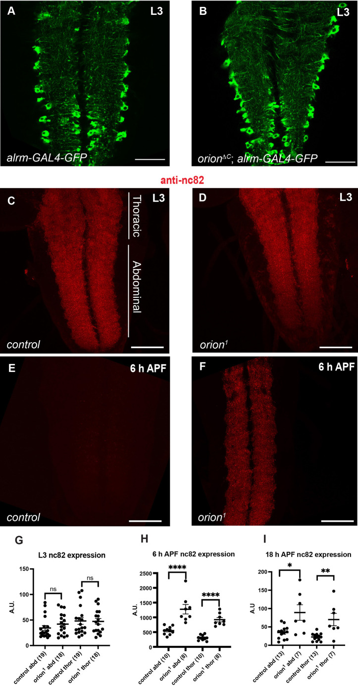
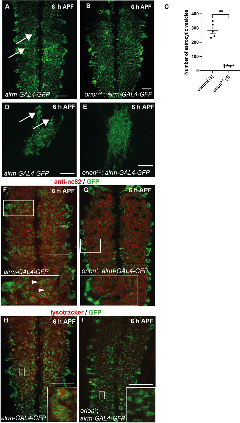
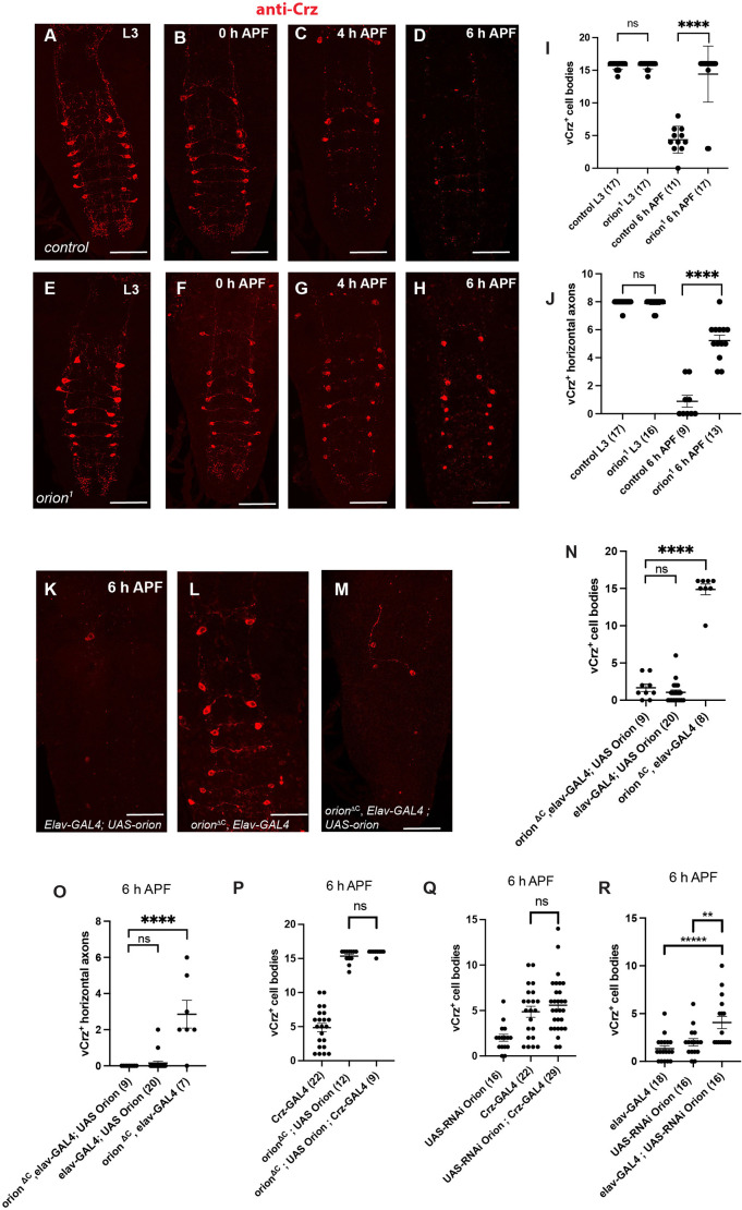
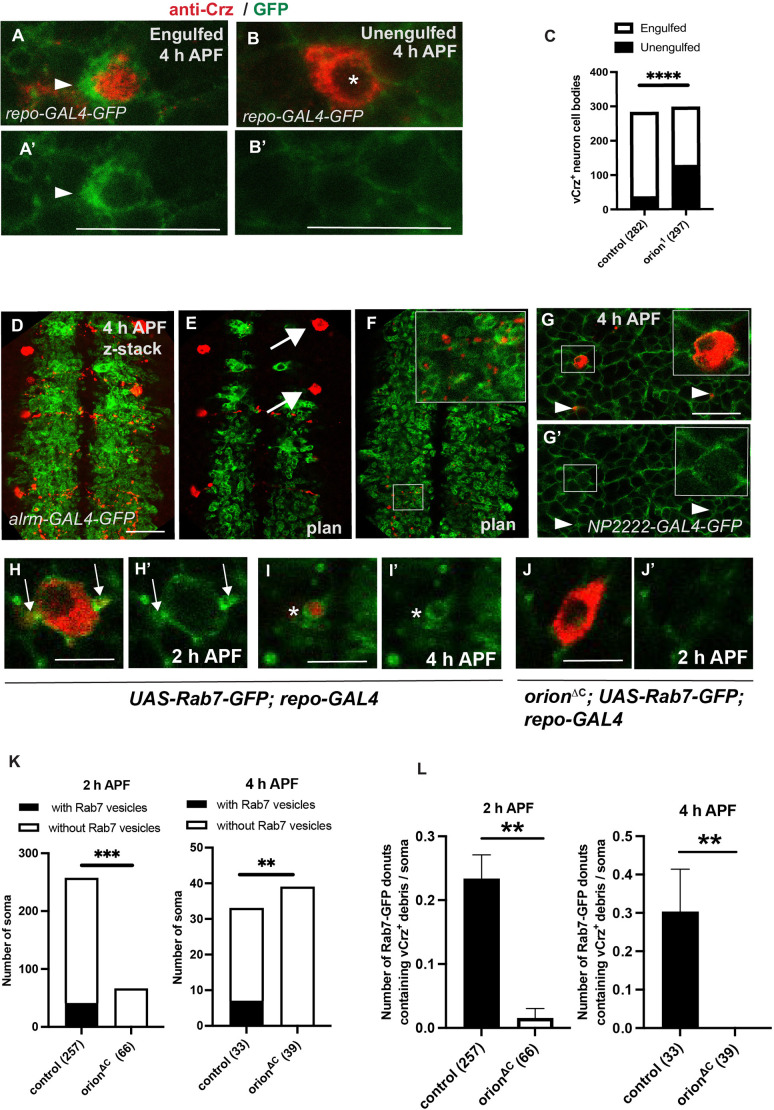
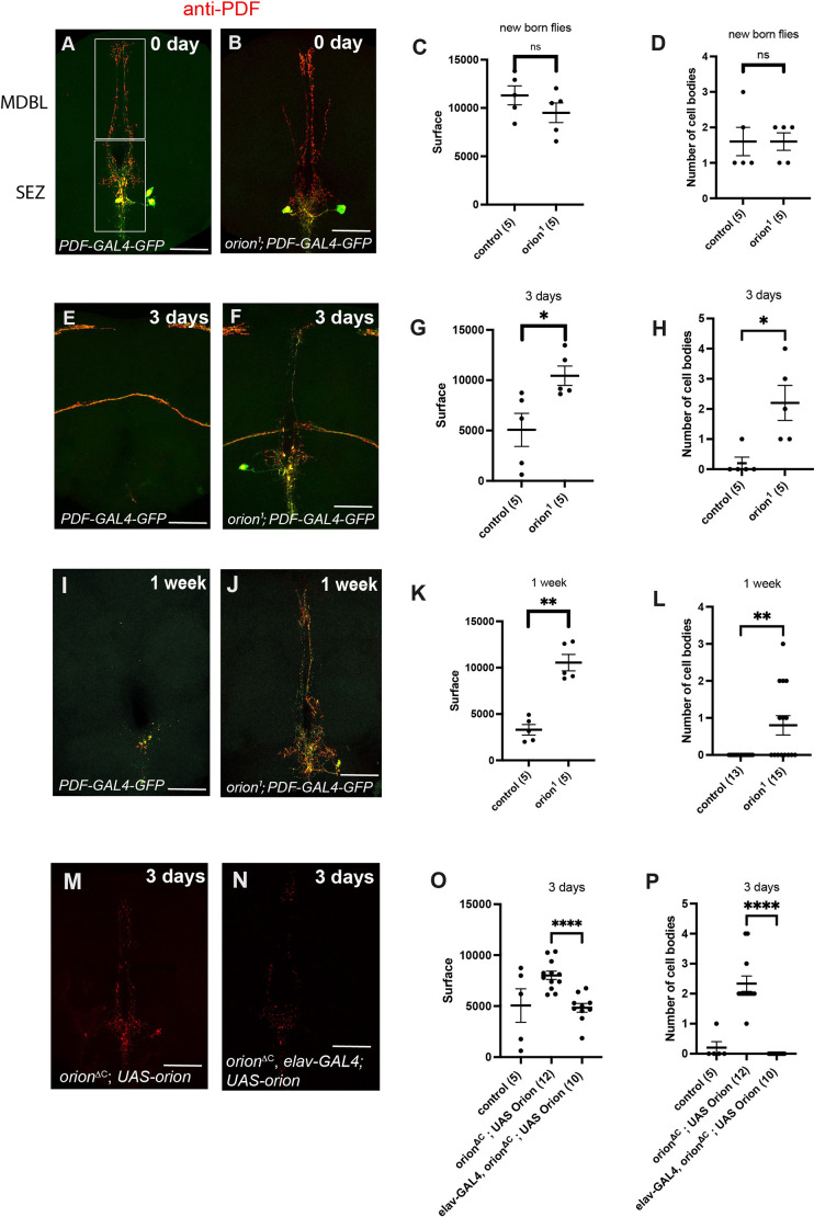
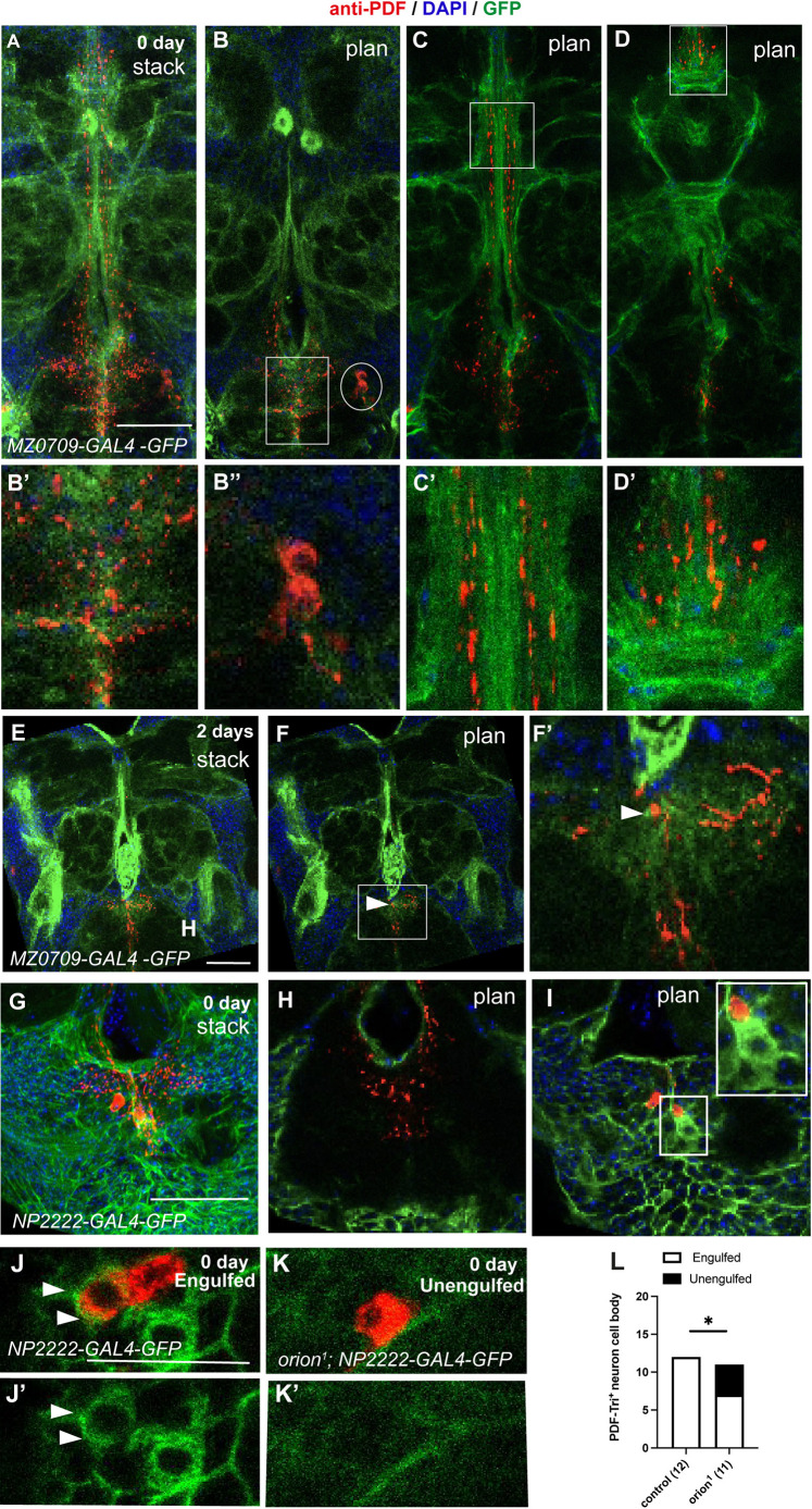
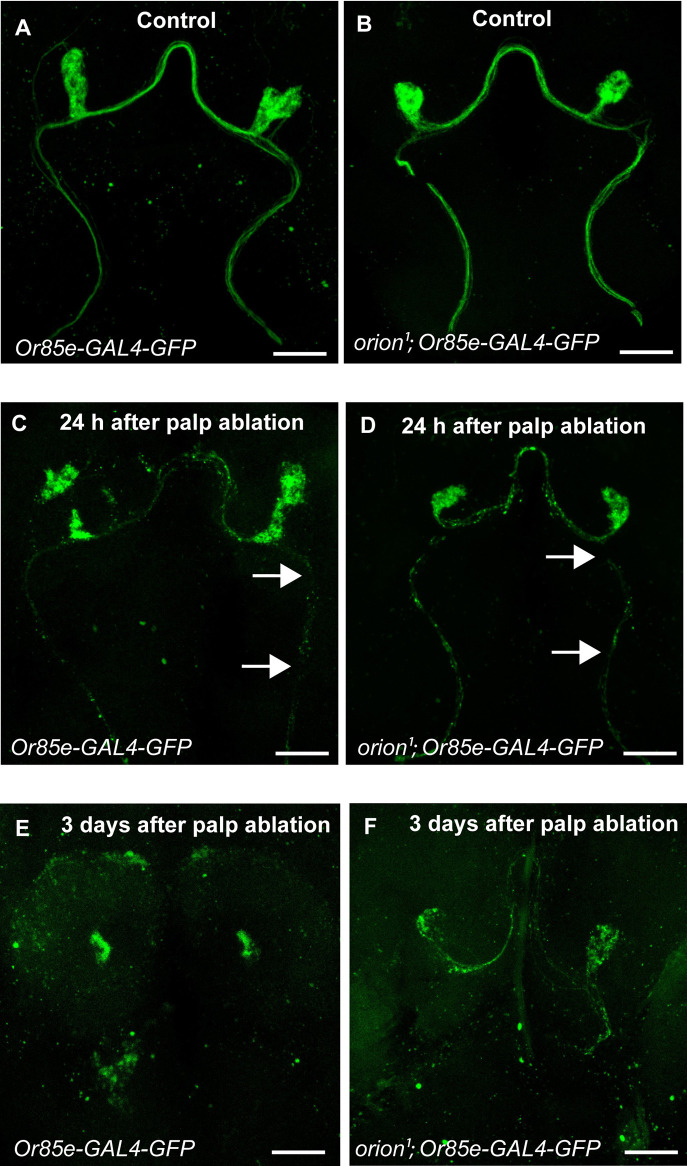
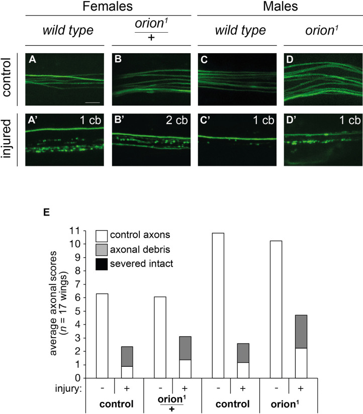
During animal development, neurons often form exuberant or inappropriate axons and dendrites at early stages, followed by the refinement of neuronal circuits at late stages. Neural circuit refinement leads to the production of neuronal debris in the form of neuronal cell corpses, fragmented axons and dendrites, and pruned synapses requiring disposal. Glial cells act as predominant phagocytes during neuronal remodeling and degeneration, and crucial signaling pathways between neurons and glia are necessary for the execution of phagocytosis. Chemokine-like mushroom body neuron-secreted Orion is essential for astrocyte infiltration into the γ axon bundle leading to γ axon pruning. Here, we show a role of Orion in debris engulfment and phagocytosis in Drosophila. Interestingly, Orion is involved in the overall transformation of astrocytes into phagocytes. In addition, analysis of several neuronal paradigms demonstrates the role of Orion in eliminating both peptidergic vCrz+ and PDF-Tri neurons via additional phagocytic glial cells like cortex and/or ensheathing glia. Our results suggest that Orion is essential for phagocytic activation of astrocytes, cortex and ensheathing glia, and point to Orion as a trigger of glial infiltration, engulfment and phagocytosis.

Keywords: Drosophila; Axon and cell body remodeling; Chemokine-like Orion; Glia; Neuron; Phagocytosis.


Conflict of interest statement

Competing interests The authors declare no competing or financial interests.

Figures

Fig. 1.
Fig. 1.
Orion is required for the elimination of synaptic material in the astrocyte-infiltrated VNC. (A,B) Astrocytes in controls (A) and orionΔC (B) are visualized by the expression of alrm-GAL4-driven UAS-mCD8-GFP (green) at larval stage (n≥20 for each condition). Note that astrocyte extensions infiltrate the larval neuropil in both conditions. (C-F) Active zones were labeled with the nc82 antibody (antibody for Brp, red) at larval (C,D) and 6 h APF (E,F) stages in control (C,E) and orion1 mutants (D,F) (n≥20 for each condition). Note the higher amount of nc82 staining observed at 6 h APF in F compared with wild type in E. Confocal images are z-projections. (G-I) Quantifications of expression levels of nc82+ puncta in arbitrary units at L3 (G), 6 h APF (H) and 18 h APF (I) in thoracic (thor) and abdominal (abd) regions (shown in C). Genotypes are listed in supplementary Materials and Methods, List of fly strains. Results are mean±s.e.m. n values are indicated in a parenthesis for each condition. Replicated three times. *P<0.05, **P<0.01, ****P<0.0001 (Mann–Whitney U-test). ns, non-significant. See Table S1 for raw data. Scale bars: 40 µm in A,B; 50 µm in C-F.
Fig. 2.
Fig. 2.
Orion regulates the transformation of astrocytes into phagocytes that engulf and phagocytose synaptic material in the pupal neuropil. (A-I) Astrocytes are visualized by the expression of alrm-GAL4-driven UAS-mCD8-GFP (green) in VNCs (A,B,F-I) and astrocyte clones (D,E). Red staining represents either active zones labeled with the nc82 antibody (F,G) or phagosomes labeled with a lysotracker staining (H,I). GFP-labeled vesicular structures are shown by arrows in A,D. Vesicles are mostly not observed, or are very small in orion mutants (B,E). n≥20 VNC and n≥10 clones in controls and orion mutants. (C) Quantification of the number of astrocytic vesicles in control and orion mutant whole VNC. The number of VNC analyzed is included in a parenthesis for each condition. Results are mean±s.e.m. **P<0.01 (Mann–Whitney U-test). Insets in F show astrocytic vesicles containing engulfed synaptic debris (arrowheads), absent in orion mutants (G). Inset in H shows astrocytic vesicles containing acidic phagosomes labeled with lysotracker. Note that lysotracker staining is not observed in orion1 vesicles (I). Confocal images are z-projections. Genotypes are listed in supplementary Materials and Methods, List of fly strains. Replicated three times. See Table S2 for raw data. Scale bars: 30 µm in A,B; 20 µm in D,E; 40 µm in F-I.
Fig. 3.
Fig. 3.
Orion is required for the elimination of vCrz+ cell bodies and neurites. (A-J) vCrz+ neurons were labeled with an anti-Crz antibody (red) at the indicated time points at larval (L3) and pupal stages in controls (A-D) and orion1 mutants (E-H). (I) Quantification of vCrz+ cell bodies. (J) Quantification of vCrz+ horizontal axons. (K-M) 6 h APF vCrz+ neurons were labeled with anti-Crz (red) in controls (K) and orion mutants (L). Neuronal expression of orion rescued the orionΔC phenotype (M). (N-R) Quantification of vCrz+ cell bodies (N,P-R) and horizontal axons (O) in rescue experiments using UAS-orion driven by elav-GAL4 (N,O) or Crz-GAL4 (P) and quantification of vCrz+ cell bodies in experiments using UAS-Orion-RNAi driven by Crz-GAL4 (Q) and elav-GAL4 (R). Genotypes are listed in supplementary Materials and Methods, List of fly strains. Error bars represent mean±s.e.m. n values are indicated in a parenthesis for each condition. **P<0.01, ****P<0.0001 (Mann–Whitney U-test). ns, non-significant. Replicated at least twice. See Table S3 for raw data. Scale bars: 70 µm in A-H; 40 µm in K-M.
Fig. 4.
Fig. 4.
The elimination of vCrz+ neurites and cell bodies is mediated by astrocytes and cortex glia respectively. (A,B) Confocal z-stacks (from five to seven plans) showing expression of UAS-mCD8-GFP under the control of repo-GAL4 (green) and anti-Crz staining (red) in 4 h APF neurons. Apoptotic cell body enwrapped in extended GFP+ cortex glia forming a phagocytic cup is shown (arrowheads) in A. A′ shows a single channel of A. Unengulfed cell body (B) displays discernible and uncondensed nuclear morphology (asterisk). GFP+ cortex glia still reaches the cell body but with very thin processes (B′, single channel of B). (C) Quantification of vCrz+ cell bodies engulfed and unengulfed by cortex glia in control versus orion mutant is shown. n values are indicated in a parenthesis for each condition. (D-F) Astrocytic glia visualized by the expression of alrm-GAL4-driven UAS-mCD8-GFP (green) and vCrz+ neurons by an anti-Crz antibody (red) at 4 h APF. (E) Arrows point to vCrz+ cell bodies which are not reached by astrocytic extensions. (F) Astrocyte extensions engulf vCrz+ debris. See inset for higher magnification. D is a z-projection confocal image, E and F are single confocal plans from D. (G,G′) Confocal plans showing cortex glia visualized by the expression of NP2222-GAL4-driven UAS-mCD8-GFP (green) and vCrz+ neurons by an anti-Crz antibody (red) at 4 h APF. Insets illustrate a vCrz+ cell body reached by cortex glia. Engulfed debris are labeled by arrowheads. n>5. (H-J′) are confocal plans showing UAS-Rab7-GFP driven by repo-GAL4 (green) and vCrz+ neurons labeled by an anti-Crz antibody (red) at 2 h and 4 h APF in wild type (H,I) and orion mutants (J). Rab7-GFP donuts attached to the soma contain vCrz+ soma-derived debris and are indicated by an arrow in H and H′. Individual donuts containing Crz+ debris are labeled by an asterisk in I,I′. Note the absence of Rab7 vesicles in orion mutants (J). H′,I′ and J′ show a single channel of H, I and J, respectively. (K,L) Quantification of the number of somas with or without Rab7 vesicles (K) and quantification of Rab7-GFP donuts containing vCrz+ debris per soma (L) at 2 h and 4 h APF. **P<0.01, ***P<0.001; ****P<0.0001 (Chi-square test). n values are indicated in a parenthesis for each condition and represent a number of soma (see raw data for number of animals). Replicated at least twice. Genotypes are listed in supplementary Materials and Methods, List of fly strains. See Table S4 for raw data. Scale bars: 20 µm in A,B; 30 µm in D-G; 10 µm in H-J.
Fig. 5.
Fig. 5.
Orion mutants retain developmentally transient PDF-Tri neurons. (A,B,E,F,I,J) Confocal z-stacks showing PDF-Tri neurons visualized by the expression of PDF-GAL4-driven UAS-mCD8-GFP (green) and labeled with anti-PDF antibody (red) at the indicated time points in controls (A,E,I) and orion1 mutants (B,F,J). (C,G,K) Surface (in µm2) occupied by the PDF-Tri arborization at 0 days (new born flies), 3 days and 1 week, respectively, in controls and orion mutants. (D,H,L) PDF-Tri cell bodies at 0 day, 3 days and 1 week in controls and orion mutants. (M,N) Confocal z-stacks showing PDF-Tri neurons labeled with an anti-PDF antibody at 3 days after birth in orionΔC mutants (M) and in orionΔC rescued by the expression of elav-GAL4-driven UAS-orion (N). (O,P) Rescue quantifications of PDF-Tri surface (in µm2) (O) and number of cell bodies (P) are shown. Genotypes are listed in supplementary Materials and Methods, List of fly strains. Error bars represent mean±s.e.m. *P<0.05, **P<0.01, ****P<0.0001 (Mann–Whitney U-test). ns, non-significant. n values are indicated in a parenthesis for each condition. Replicated at least twice. See Table S5 for raw data. Scale bars: 50 µm.
Fig. 6.
Fig. 6.
Orion mediates PDF-Tri neuron elimination via cortex and ensheathing glia. (A-F′) Confocal z-stacks or plans (labeled stack or plan) showing ensheathing glia visualized by the expression of MZ0709-GAL4-driven UAS-mCD8-GFP (green) and PDF-Tri neurons labeled with anti-PDF antibody (red) and DAPI (blue) at 0 (A) and 2 (E) days. Different regions of the brain contained in these z-stacks are shown as single confocal plans (B-D′ for z-stack in A, F,F′ for z-stack in E). Suboesophageal zone (SEZ) region is included in a rectangle in B and F, distal medial bundle (MDBL) regions are included in a rectangle in C and D. These regions are shown at higher magnification in B′,C′,D′ and F′, respectively, showing PDF-Tri neurites engulfed by ensheathing glia. Arrowheads point to ensheathing glia-engulfed debris in F and F′. Oval in B encircles a PDF-Tri cell body devoid of ensheathing glia, which does not engulf PDF-Tri cell bodies (higher magnification is in B″). (G) Confocal z-stack labeled with anti-PDF antibody (red) and DAPI (blue) and showing cortex glia visualized by the expression of NP2222-GAL4-driven UAS-mCD8-GFP (green) at 0 day. (H,I) Single confocal plans showing PDF-Tri dendrites (H) and cell bodies (I) contained in the panel G image stack. Note the absence of PDF-Tri dendrite surrounded by cortex glia in H and the high amount of cortex glia extensions present around the PDF-Tri cell bodies in I. A cortex glia cell phagocytosing a PDF-Tri cell body (red) is included in the region enclosed by the square in I. n≥5 for each condition. Replicated twice. (J-K′) Confocal plans showing expression of UAS-mCD8-GFP under the control of NP2222-GAL4 (green) and anti-PDF staining (red) in day 0 neurons. An engulfed apoptotic PDF-Tri cell body enwrapped in thick GFP+ cortex glia extensions is indicated with arrowheads in J and J′. An unengulfed PDF-Tri cell body is shown in K. J′ and K′ show a single channel of J and K, respectively. (L) Quantification of PDF-Tri+ cell bodies engulfed and unengulfed by cortex glia is shown. n values are indicated in a parenthesis for each condition. Statistically significant differences were observed between the two groups (*P<0.05, Fisher test). Genotypes are listed in supplementary Materials and Methods, List of fly strains. See Table S6 for raw data. Scale bars: 50 µm in A-I; 20 µm in J,K.
Fig. 7.
Fig. 7.
Orion is not involved in the elimination of ORN debris by ensheathing glia after injury by palp ablation. (A,B) Confocal z-stacks showing ORNs before injury (control) visualized by the expression of Or85e-GAL4-driven UAS-mCD8-GFP (green) in wild type (A) and orion1 mutants (B). (C-F) Confocal z-stacks showing ORNs 24 h after palp ablation visualized by the expression of Or85e-GAL4-driven UAS-mCD8-GFP (green) in wild type (C) and orion1 mutants (D) and 3 days after palp ablation in wild type (E) and orion1 mutants (F). Note the axon discontinuities observed in both conditions in C and D, 24 h after palp ablation (arrows), and that only disseminated debris are observed in both conditions 3 days after palp ablation. Genotypes are listed in supplementary Materials and Methods, List of fly strains. n≥20 brains for each condition. Replicated twice. Scale bars: 30 µm.
Fig. 8.
Fig. 8.
Orion is dispensable for clearing axonal debris after axotomy in the wing. (A-D′) Images are confocal z-stacks showing L1 vein axons labeled by dpr1-GAL4-driven UAS-mCD8-GFP (green). Representative pictures of control and injured (7 dpa) axons in wild type, heterozygous orion1 and hemizygous orion1 animals are shown. In the upper right corner of each image, the number of neuronal cell bodies (cb) indicates the number of uninjured, thus attached, neurons and therefore the expected intact axons in the field of view. (E) Quantification of average axonal scores of uninjured controls, debris and severed intact axons (white, gray and black, respectively). MARCM clones were generated in animals of the indicated genetic background. Genotypes are listed in supplementary Materials and Methods, List of fly strains. See Table S7 for raw data. Scale bar: 5 µm.

Similar articles

Cited by

References

    1. Arnoux, I. and Audinat, E. (2015). Fractalkine signaling and microglia functions in the developing brain. Neural Plast. 2015, 689404. 10.1155/2015/689404 - DOI - PMC - PubMed
    1. Awasaki, T. and Ito, K. (2004). Engulfing action of glial cells is required for programmed axon pruning during Drosophila metamorphosis. Curr. Biol. 14, 668-677. 10.1016/j.cub.2004.04.001 - DOI - PubMed
    1. Bittern, J., Pogodalla, N., Ohm, H., Brüser, L., Kottmeier, R., Schirmeier, S. and Klämbt, C. (2021). Neuron-glia interaction in the Drosophila nervous system. Dev. Neurobiol. 81, 438-452. 10.1002/dneu.22737 - DOI - PubMed
    1. Boulanger, A. and Dura, J.-M. (2015). Nuclear receptors and Drosophila neuronal remodeling. Biochim. Biophys. Acta 1849, 187-195. 10.1016/j.bbagrm.2014.05.024 - DOI - PubMed
    1. Boulanger, A. and Dura, J. M. (2022). Neuron-glia crosstalk in neuronal remodeling and degeneration: neuronal signals inducing glial cell phagocytic transformation in Drosophila. BioEssays 44, e2100254. 10.1002/bies.202100254 - DOI - PubMed

LinkOut - more resources