eNOS Regulates Lymphatic Valve Specification by Controlling β-Catenin Signaling During Embryogenesis in Mice
- PMID: 37767708
- PMCID: PMC10655861
- DOI: 10.1161/ATVBAHA.123.319405
eNOS Regulates Lymphatic Valve Specification by Controlling β-Catenin Signaling During Embryogenesis in Mice
Abstract
Background: Lymphatic valves play a critical role in ensuring unidirectional lymph transport. Loss of lymphatic valves or dysfunctional valves are associated with several diseases including lymphedema, lymphatic malformations, obesity, and ileitis. Lymphatic valves first develop during embryogenesis in response to mechanotransduction signaling pathways triggered by oscillatory lymph flow. In blood vessels, eNOS (endothelial NO synthase; gene name: Nos3) is a well-characterized shear stress signaling effector, but its role in lymphatic valve development remains unexplored.
Methods: We used global Nos3-/- mice and cultured human dermal lymphatic endothelial cells to investigate the role of eNOS in lymphatic valve development, which requires oscillatory shear stress signaling.
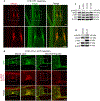
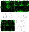
Results: Our data reveal a 45% reduction in lymphatic valve specification cell clusters and that loss of eNOS protein inhibited activation of β-catenin and its nuclear translocation. Genetic knockout or knockdown of eNOS led to downregulation of β-catenin target proteins in vivo and in vitro. However, pharmacological inhibition of NO production did not reproduce these effects. Co-immunoprecipitation and proximity ligation assays reveal that eNOS directly binds to β-catenin and their binding is enhanced by oscillatory shear stress. Finally, genetic ablation of the Foxo1 gene enhanced FOXC2 expression and partially rescued the loss of valve specification in the eNOS knockouts.
Conclusions: In conclusion, we demonstrate a novel, NO-independent role for eNOS in regulating lymphatic valve specification and propose a mechanism by which eNOS directly binds β-catenin to regulate its nuclear translocation and thereby transcriptional activity.
Keywords: cadherins; catenins; lymphedema; mechanotransduction; nitric oxide.
Conflict of interest statement
Figures


Update of
-
Endothelial Nitric Oxide Synthase Regulates Lymphatic Valve Specification By Controlling β - catenin Signaling During Embryogenesis.bioRxiv [Preprint]. 2023 Apr 11:2023.04.10.536303. doi: 10.1101/2023.04.10.536303. bioRxiv. 2023. Update in: Arterioscler Thromb Vasc Biol. 2023 Nov;43(11):2197-2212. doi: 10.1161/ATVBAHA.123.319405. PMID: 37090551 Free PMC article. Updated. Preprint.
Similar articles
-
Endothelial Nitric Oxide Synthase Regulates Lymphatic Valve Specification By Controlling β - catenin Signaling During Embryogenesis.bioRxiv [Preprint]. 2023 Apr 11:2023.04.10.536303. doi: 10.1101/2023.04.10.536303. bioRxiv. 2023. Update in: Arterioscler Thromb Vasc Biol. 2023 Nov;43(11):2197-2212. doi: 10.1161/ATVBAHA.123.319405. PMID: 37090551 Free PMC article. Updated. Preprint.
-
Rictor, an mTORC2 Protein, Regulates Murine Lymphatic Valve Formation Through the AKT-FOXO1 Signaling.Arterioscler Thromb Vasc Biol. 2024 Sep;44(9):2004-2023. doi: 10.1161/ATVBAHA.124.321164. Epub 2024 Aug 1. Arterioscler Thromb Vasc Biol. 2024. PMID: 39087350 Free PMC article.
-
Mechanotransduction activates canonical Wnt/β-catenin signaling to promote lymphatic vascular patterning and the development of lymphatic and lymphovenous valves.Genes Dev. 2016 Jun 15;30(12):1454-69. doi: 10.1101/gad.282400.116. Epub 2016 Jun 16. Genes Dev. 2016. PMID: 27313318 Free PMC article.
-
Mechanosensation and Mechanotransduction by Lymphatic Endothelial Cells Act as Important Regulators of Lymphatic Development and Function.Int J Mol Sci. 2021 Apr 12;22(8):3955. doi: 10.3390/ijms22083955. Int J Mol Sci. 2021. PMID: 33921229 Free PMC article. Review.
-
Interplay of mechanotransduction, FOXC2, connexins, and calcineurin signaling in lymphatic valve formation.Adv Anat Embryol Cell Biol. 2014;214:67-80. doi: 10.1007/978-3-7091-1646-3_6. Adv Anat Embryol Cell Biol. 2014. PMID: 24276887 Review.
Cited by
-
Zmiz1 is a novel regulator of lymphatic endothelial cell gene expression and function.bioRxiv [Preprint]. 2023 Jul 22:2023.07.22.550165. doi: 10.1101/2023.07.22.550165. bioRxiv. 2023. Update in: PLoS One. 2024 May 8;19(5):e0302926. doi: 10.1371/journal.pone.0302926. PMID: 37503058 Free PMC article. Updated. Preprint.
-
A neuro-lymphatic communication guides lymphatic development by CXCL12 and CXCR4 signaling.Development. 2024 Nov 15;151(22):dev202901. doi: 10.1242/dev.202901. Epub 2024 Nov 26. Development. 2024. PMID: 39470100
-
Transplantation of engineered vascularized lymphatic tissue using LEC and in vivo AV loop model to enhance lymphangiogenesis and restore lymphatic drainage in a lymphadenectomy rat model.J Tissue Eng. 2025 Aug 10;16:20417314251360755. doi: 10.1177/20417314251360755. eCollection 2025 Jan-Dec. J Tissue Eng. 2025. PMID: 40792201 Free PMC article.
-
Zmiz1 is a novel regulator of lymphatic endothelial cell gene expression and function.PLoS One. 2024 May 8;19(5):e0302926. doi: 10.1371/journal.pone.0302926. eCollection 2024. PLoS One. 2024. PMID: 38718095 Free PMC article.
References
-
- Czepielewski RS, Erlich EC, Onufer EJ, Young S, Saunders BT, Han YH, Wohltmann M, Wang PL, Kim KW, Kumar S, et al. Ileitis-associated tertiary lymphoid organs arise at lymphatic valves and impede mesenteric lymph flow in response to tumor necrosis factor. Immunity. 2021;54:2795–2811.e2799. doi: 10.1016/j.immuni.2021.10.003 - DOI - PMC - PubMed
Publication types
MeSH terms
Substances
Grants and funding
LinkOut - more resources
Full Text Sources
Molecular Biology Databases
Research Materials
Miscellaneous

