. 2023 Sep 28;11(5):e0130123.
doi: 10.1128/spectrum.01301-23. Online ahead of print.

The plant pathogenic bacterium Candidatus Liberibacter solanacearum induces calcium-regulated autophagy in midgut cells of its insect vector Bactericera trigonica

Affiliations

The plant pathogenic bacterium Candidatus Liberibacter solanacearum induces calcium-regulated autophagy in midgut cells of its insect vector Bactericera trigonica

Poulami Sarkar et al. Microbiol Spectr. .

Abstract

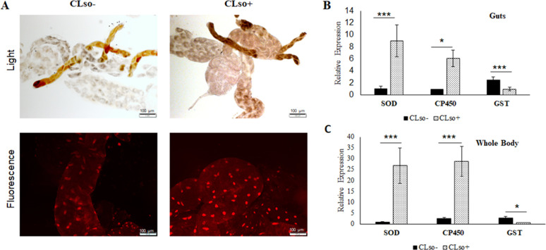
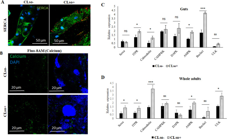
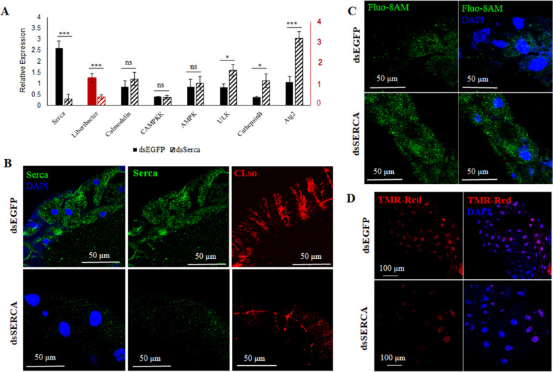
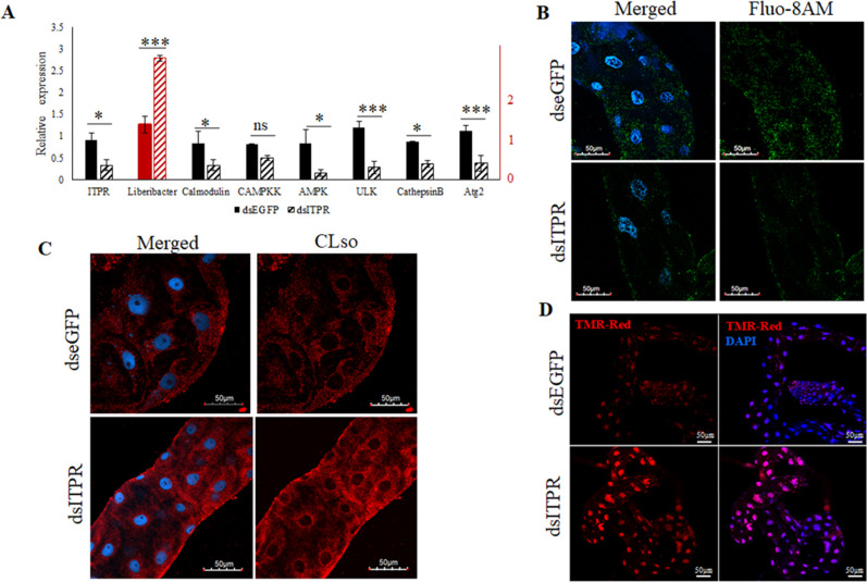
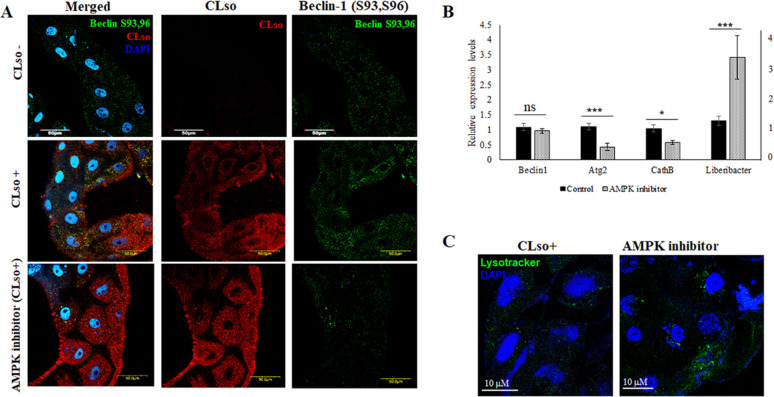
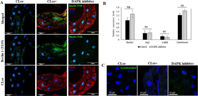
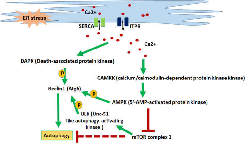
Autophagy plays an important role against pathogen infection in many organisms; however, little has been done with regard to vector-borne plant and animal pathogens, that sometimes replicate and cause deleterious effects in their vectors. Candidatus Liberibacter solanacearum (CLso) is a fastidious gram-negative phloem-restricted plant pathogen and vectored by the carrot psyllid, Bactericera trigonica. The plant disease caused by this bacterium is called carrot yellows and has recently gained much importance due to worldwide excessive economical losses. Here, we demonstrate that calcium ATPase, cytosolic calcium, and most importantly Beclin-1 have a role in regulating autophagy and its association with Liberibacter inside the psyllid. The presence of CLso generates reactive oxygen species and induces the expression of detoxification enzymes in the psyllid midguts, a main site for bacteria transmission. CLso also induces the expression of both sarco/endoplasmic reticulum Ca2+pump (SERCA) and 1,4,5-trisphosphate receptors (ITPR) in midguts, resulting in high levels of calcium in the cellular cytosol. Silencing these genes individually disrupted the calcium levels in the cytosol and resulted in direct effects on autophagy and subsequently on Liberibacter persistence and transmission. Inhibiting Beclin1-phosphorylation through different calcium-induced kinases altered the expression of autophagy and CLso titers and persistence. Based on our results obtained from the midgut, we suggest the existence of a direct correlation between cytosolic calcium levels, autophagy, and CLso persistence and transmission by the carrot psyllid. IMPORTANCE Plant diseases caused by vector-borne Liberibacter species are responsible for the most important economic losses in many agricultural sectors. Preventing these diseases relies mostly on chemical sprays against the insect vectors. Knowledge-based interference with the bacteria-vector interaction remains a promising approach as a sustainable solution. For unravelling how Liberibacter exploits molecular pathways in its insect vector for transmission, here, we show that the bacterium manipulates calcium levels on both sides of the endoplasmic reticulum membrane, resulting in manipulating autophagy. Silencing genes associated with these pathways disrupted the calcium levels in the cytosol and resulted in direct effects on autophagy and Liberibacter transmission. These results demonstrate major pathways that could be exploited for manipulating and controlling the disease transmission.

Keywords: Liberibacter; SERCA; autophagy; calcium signaling; psyllid.


Conflict of interest statement

The authors declare no conflict of interest.

Figures

Fig 1
Fig 1
ROS detection in CLso-infected psyllid midguts. (A) Light and fluorescence detection of ROS using DHE stain. (B) Real-time PCR analysis for SOD, CP450, and GST in guts and (C) in whole psyllids. * indicates P ≤ 0.05 and *** indicate P ≤ 0.01. Error bars denote SE with n ≥ 10.
Fig 2
Fig 2
Differential expressions of SERCA, calcium, and calcium-signaling genes. (A) Immunostaining of SERCA (green) and CLso (red) in CLso-free (CLso–) and CLso-infected (CLso+) psyllid midguts counterstained with DAPI (blue). (B) Detection of cytosolic calcium levels (green) using Fluo-8AM staining in CLso– and CLso+ psyllid midguts, counterstained with DAPI (blue). (C) Real-time PCR analysis for the expression of SERCA, ITPR, and calcium-signaling cascade genes in the midguts and (D) in whole psyllids. * indicates P ≤ 0.05, *** indicates P ≤ 0.01, and ns indicates not significant. Error bars denote SE with n ≥ 10.
Fig 3
Fig 3
Effect of silencing SERCA on CLso and autophagy. (A) Real-time PCR analysis of the differential expression change in SERCA and corresponding calcium-signaling genes along with CLso abundance in the psyllid midguts following SERCA silencing. * denotes P ≤ 0.05, *** indicates P ≤ 0.01, and ns indicates not significant. Error bars denote SE with n ≥ 10. (B) Representative image of immunostaining analysis of SERCA (green) and CLso (red) in the midguts upon SERCA silencing, counterstained with DAPI (blue). (C) Elevated levels of cytosolic calcium (green) after silencing SERCA in midguts. (D) TUNEL assay showing reduced apoptosis in SERCA silenced midguts.
Fig 4
Fig 4
Effect of ITPR silencing on CLso and autophagy. (A) Real-time PCR analysis showing differential expression levels of ITPR, calcium-signaling genes, and CLso abundance in the psyllid midguts following silencing ITPR. * denotes P ≤ 0.05, *** denotes P ≤ 0.01, and ns indicates not significant. Error bars denote SE with n ≥ 10. (B) Fluo-8AM staining of cytosolic calcium (green) after ITPR silencing and counterstaining with DAPI (blue). (C) Immunostaining of CLso (red) in the psyllid midguts following ITPR Silencing. (D) Increased apoptosis in ITPR-silenced midguts as detected by TUNEL assay using TMR-red (red) and DAPI (blue).
Fig 5
Fig 5
Effect of AMPK inhibitor on Beclin1 phosphorylation, autophagy, and CLso. (A) Immunostaining analysis reveals reduced Beclin1 phosphorylation (green) at Ser93,96 sites and higher accumulation of CLso (red) in the AMPK-inhibited psyllid midguts, counterstained with DAPI (blue). (B) Real-time PCR assay for the differential expression of Beclin1, autophagy genes, and CLso abundance following AMPK inhibition. P ≤ 0.05 is indicated by *, P ≤ 0.01 by ***, and ns indicates not significant. Error bars denote SE with n ≥ 15. (C) Autolysosome detection using Lysotracker DND (green) counterstained with DAPI (blue).
Fig 6
Fig 6
Effect of DAPK inhibitor on Beclin phosphorylation, autophagy and CLso abundance. (A) Immunostaining analysis for Beclin1 phosphorylation at site Thr119 (green) and CLso (red) counterstained with DAPI (blue). (B) Real-time analysis showing differential expression levels for Beclin1, autophagy genes and CLso titer. ns denotes not significant. Error bars denote SE with n ≥ 15. (C) Autolysosome detection using Lysotracker DND (green) counterstained with DAPI (blue).
Fig 7
Fig 7
Depiction of calcium signaling cascade leading to autophagy. Calcium influx (SERCA) pumps and efflux (ITPR) pumps maintain calcium homeostasis in the ER as well as in the cytosol. ER stress causes elevated cytosolic calcium levels, which in turn activates downstream protein kinases leading to the activation of Beclin1 which helps in the initiation of autophagosome formation leading to autophagy.

Similar articles

Cited by

References

    1. Bartolome A, Guillen C, Benito M. 2012. Autophagy plays a protective role in endoplasmic reticulum stress-mediated pancreatic β cell death. Autophagy 8:1757–1768. doi:10.4161/auto.21994 - DOI - PMC - PubMed
    1. Parzych KR, Klionsky DJ. 2014. An overview of autophagy: morphology, mechanism, and regulation. Antioxid Redox Signal 20:460–473. doi:10.1089/ars.2013.5371 - DOI - PMC - PubMed
    1. Niu M, Dai X, Zou W, Yu X, Teng W, Chen Q, Sun X, Yu W, Ma H, Liu P. 2017. Autophagy, endoplasmic reticulum stress and the unfolded protein response in intracerebral hemorrhage. Transl Neurosci 8:37–48. doi:10.1515/tnsci-2017-0008 - DOI - PMC - PubMed
    1. Thorburn A. 2008. Apoptosis and autophagy: regulatory connections between two supposedly different processes. Apoptosis 13:1–9. doi:10.1007/s10495-007-0154-9 - DOI - PMC - PubMed
    1. Wang Y, Qin Z. 2013. Coordination of autophagy with other cellular activities. Acta Pharmacol Sin 34:585–594. doi:10.1038/aps.2012.194 - DOI - PMC - PubMed

LinkOut - more resources