. 2023 Dec;43(1):1-10.
doi: 10.1080/01652176.2023.2265172. Epub 2023 Oct 9.

Domestic cat hepadnavirus detection in blood and tissue samples of cats with lymphoma

Affiliations

Domestic cat hepadnavirus detection in blood and tissue samples of cats with lymphoma

Chutchai Piewbang et al. Vet Q. 2023 Dec.

Abstract

Domestic cat hepadnavirus (DCH), a relative hepatitis B virus (HBV) in human, has been recently identified in cats; however, association of DCH infection with lymphoma in cats is not investigated. To determine the association between DCH infection and feline lymphoma, seven hundred and seventeen cats included 131 cats with lymphoma (68 blood and 63 tumor samples) and 586 (526 blood and 60 lymph node samples) cats without lymphoma. DCH DNA was investigated in blood and formalin-fixed paraffin-embedded (FFPE) tissues by quantitative polymerase chain reaction (qPCR). The FFPE lymphoma tissues were immunohistochemically subtyped, and the localization of DCH in lymphoma sections was investigated using in situ hybridization (ISH). Feline retroviral infection was investigated in the DCH-positive cases. DCH DNA was detected in 16.18% (11/68) (p = 0.002; odds ratio [OR], 5.15; 95% confidence interval [CI], 2.33-11.36) of blood and 9.52% (6/63) (p = 0.028; OR, 13.68; 95% CI, 0.75-248.36) of neoplastic samples obtained from lymphoma cats, whereas only 3.61% (19/526) of blood obtained from non-lymphoma cats was positive for DCH detection. Within the DCH-positive lymphoma, in 3/6 cats, feline leukemia virus was co-detected, and in 6/6 were B-cell lymphoma (p > 0.9; OR, 1.93; 95% CI, 0.09-37.89) and were multicentric form (p = 0.008; OR, 1.327; 95% CI, 0.06-31.18). DCH was found in the CD79-positive pleomorphic cells. Cats with lymphoma were more likely to be positive for DCH than cats without lymphoma, and infection associated with lymphoma development needs further investigations.

Keywords: B-cell lymphoma; domestic cat hepadnavirus; feline; hepatitis virus; localization.


Conflict of interest statement

No potential conflict of interest was reported by the authors.

Figures
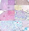
Figure 1.
Figure 1.
Histopathological features of DCH infection in lymphoma tissue of cats. (A, E-J) Sections from case no. 3. (A) The lymphoid follicles are diffusely expanded by large neoplastic lymphocytes separating coarse fibrovascular stroma. Neoplastic lymphocytes have moderate amounts of cytoplasm with distinct cellular borders. The nuclei are 2 times larger than red blood cells, round, and finely stippled, containing a prominent nucleolus (square box is indicated for the inset). (B-D) DCH qPCR-positive lymphoma section of case no.6. (B) Diffuse, generalized cytoplasmic IHC staining for CD79. (C) Prominent nuclear IHC staining for pax-5 in multiple neoplastic cells. (D) Cytoplasmic IHC staining for CD3 in some single cells in lymphoma tissue. (E) In situ hybridization (ISH) for DCH DNA staining (red precipitates) in lymphoma tissue revealed intensely intranuclear staining in diffuse round cells (square box is indicated for the inset). (F) Prominent nuclear staining for DCH hybridization in large, pleomorphic round cells. No DCH labeling was observed within the small round cells. (G) Dual DCH ISH (red precipitates) and CD79 IHC (green color) revealed marked, diffuse staining in the nucleus and cytoplasm of neoplastic cells, respectively. (H) Higher magnification reveals intense DCH labeling (red precipitates) in the nucleus of the cells that were cytoplasmically stained with CD79 IHC (green color). (I) Negative control using unrelated probe. No hybridization signals were present in the DCH qPCR-positive lymphoma section that was incubated with the FBoV-3 ISH probe. (J) Negative control using DNase-treated section. No hybridization was present in the DCH qPCR-positive lymphoma section treated with DNase prior to incubation with the DCH probe. (K) Negative control for dual ISH/IHC labeling. Cytoplasmic IHC CD79 staining of the DCH qPCR-positive lymphoma section incubated with the FBoV-3 probe. Bars indicate 180 µm for A, B, D and E; 80 µm for F; 120 µm for C, G, I, and J; and 20 µm for H and K.

References

    1. Aghazadeh M, Shi M, Barrs VR, McLuckie AJ, Lindsay SA, Jameson B, Hampson B, Holmes EC, Beatty JA.. 2018. A novel hepadnavirus identified in an immunocompromised domestic cat in Australia. Viruses. 10(5):269. doi: 10.3390/v10050269. - DOI - PMC - PubMed
    1. Beatty J. 2014. Viral causes of feline lymphoma: retroviruses and beyond. Vet J. 201(2):174–180. doi: 10.1016/j.tvjl.2014.05.026. - DOI - PubMed
    1. Bertone ER, Snyder LA, Moore AS.. 2002. Environmental tobacco smoke and risk of malignant lymphoma in pet cats. Am J Epidemiol. 156(3):268–273. doi: 10.1093/aje/kwf044. - DOI - PubMed
    1. Cao X, Wang Y, Li P, Huang W, Lu X, Lu H.. 2021. HBV reactivation during the treatment of non-Hodgkin lymphoma and management strategies. Front Oncol. 11:685706. doi: 10.3389/fonc.2021.685706. - DOI - PMC - PubMed
    1. Capozza P, Lanave G, Diakoudi G, Stasi F, Ghergo P, Ricci D, Santo G, Arena G, Grillo I, Delle Donne E, et al. 2021. A longitudinal observational study in two cats naturally-infected with hepadnavirus. Vet Microbiol. 254:108999. doi: 10.1016/j.vetmic.2021.108999. - DOI - PubMed