

Choriocapillaris Impairment Is Associated With Delayed Rod-Mediated Dark Adaptation in Age-Related Macular Degeneration
- PMID: 37768273
- PMCID: PMC10540875
- DOI: 10.1167/iovs.64.12.41
Choriocapillaris Impairment Is Associated With Delayed Rod-Mediated Dark Adaptation in Age-Related Macular Degeneration
Abstract
Purpose: Progress toward treatment and prevention of age-related macular degeneration (AMD) requires imaging end points that relate to vision. We investigated choriocapillaris flow signal deficits (FD%) and visual function in eyes of individuals aged ≥60 years, with and without AMD.
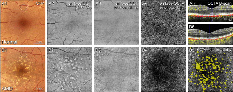
Methods: One eye of each participant in the baseline visit of the Alabama Study on Early Age-Related Macular Degeneration 2 (ALSTAR2; NCT04112667) was studied. AMD presence and severity was determined using the Age-Related Eye Disease Study (AREDS) grading system. FD% was quantified using macular spectral domain optical coherence tomography angiography (OCTA) scans. Vision tests included rod-mediated dark adaptation (RMDA), best-corrected visual acuity, and contrast sensitivity (photopic and mesopic), and microperimetric light sensitivity (scotopic, mesopic, and photopic). Presence of subretinal drusenoid deposits (SDD) was determined using multimodal imaging.
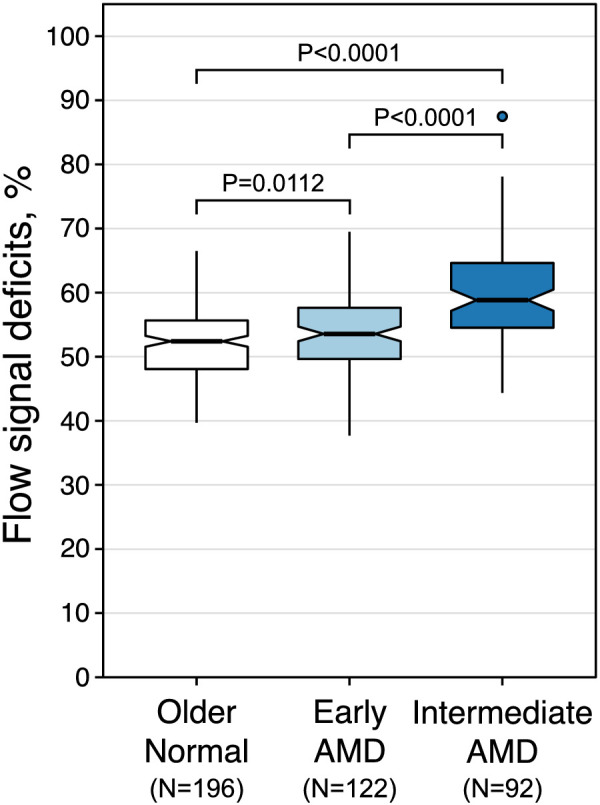
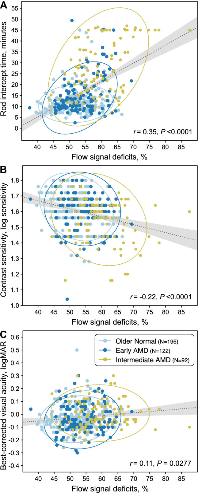
Results: In 410 study eyes of 410 participants (mean [SD] age = 71.7 years [5.9]), FD% was higher in early AMD (mean [SD] = 54.0% [5.5], N = 122) and intermediate AMD (59.8% [7.4], N = 92), compared to normal (52.1% [5.3], N = 196) eyes. Among visual functions evaluated, RMDA showed the strongest association with FD% (r = 0.35, P < 0.0001), followed by contrast sensitivity (r = -0.22, P < 0.0001). Eyes with SDD had worse FD% (58.3% [7.4], N = 87), compared to eyes without SDD (53.4% [6.0], N = 323, P = < 0.0001).
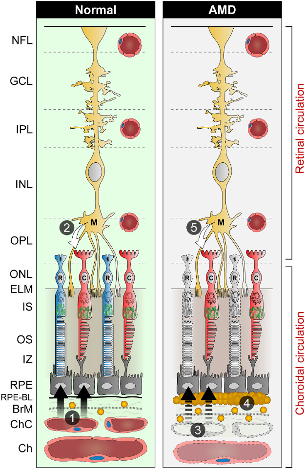
Conclusions: Choriocapillaris FD% were associated with AMD severity and with impaired vision, especially RMDA. Reduced metabolic transport and exchange across the choriocapillaris-Bruch's membrane retinal pigment epithelium (RPE) complex, a causal factor for high-risk soft drusen formation, also may impair photoreceptor sustenance from the circulation. This includes retinoid resupply, essential to dynamic rod function.
Conflict of interest statement
Disclosure:
Figures
Similar articles
-
Functionally validated imaging endpoints in the Alabama study on early age-related macular degeneration 2 (ALSTAR2): design and methods.BMC Ophthalmol. 2020 May 19;20(1):196. doi: 10.1186/s12886-020-01467-0. BMC Ophthalmol. 2020. PMID: 32429847 Free PMC article.
-
Biologically Guided Optimization of Test Target Location for Rod-mediated Dark Adaptation in Age-related Macular Degeneration: Alabama Study on Early Age-related Macular Degeneration 2 Baseline.Ophthalmol Sci. 2023 Jan 23;3(2):100274. doi: 10.1016/j.xops.2023.100274. eCollection 2023 Jun. Ophthalmol Sci. 2023. PMID: 36875335 Free PMC article.
-
How Vision Is Impaired From Aging to Early and Intermediate Age-Related Macular Degeneration: Insights From ALSTAR2 Baseline.Transl Vis Sci Technol. 2022 Jul 8;11(7):17. doi: 10.1167/tvst.11.7.17. Transl Vis Sci Technol. 2022. PMID: 35861686 Free PMC article.
-
Dark Adaptation and Its Role in Age-Related Macular Degeneration.J Clin Med. 2022 Mar 1;11(5):1358. doi: 10.3390/jcm11051358. J Clin Med. 2022. PMID: 35268448 Free PMC article. Review.
-
Perspectives on reticular pseudodrusen in age-related macular degeneration.Surv Ophthalmol. 2016 Sep-Oct;61(5):521-37. doi: 10.1016/j.survophthal.2016.02.005. Epub 2016 Mar 17. Surv Ophthalmol. 2016. PMID: 26994868 Review.
Cited by
-
Ghost vessels in the eye: Cell free choriocapillaris domains in atrophic age-related macular degeneration.Exp Eye Res. 2024 Nov;248:110128. doi: 10.1016/j.exer.2024.110128. Epub 2024 Oct 16. Exp Eye Res. 2024. PMID: 39419369
-
Dissecting the biological complexity of age-related macular degeneration: Is it one disease, multiple separate diseases, or a spectrum?Exp Eye Res. 2025 May;254:110304. doi: 10.1016/j.exer.2025.110304. Epub 2025 Feb 19. Exp Eye Res. 2025. PMID: 39983974 Free PMC article. Review.
-
Choriocapillaris Impairment, Visual Function, and Distance to Fovea in Aging and Age-Related Macular Degeneration: ALSTAR2 Baseline.Invest Ophthalmol Vis Sci. 2024 Jul 1;65(8):40. doi: 10.1167/iovs.65.8.40. Invest Ophthalmol Vis Sci. 2024. PMID: 39042400 Free PMC article.
-
Assessment of choriocapillary flow deficits in myopic children by optical coherence tomography angiography.Sci Rep. 2025 May 3;15(1):15535. doi: 10.1038/s41598-025-98579-8. Sci Rep. 2025. PMID: 40319126 Free PMC article.
-
Outer Retinal Thickness Is Associated With Cognitive Function in Normal Aging to Intermediate Age-Related Macular Degeneration.Invest Ophthalmol Vis Sci. 2024 May 1;65(5):16. doi: 10.1167/iovs.65.5.16. Invest Ophthalmol Vis Sci. 2024. PMID: 38717425 Free PMC article.
References
-
- Wong WL, Su X, Li X, et al. .. Global prevalence of age-related macular degeneration and disease burden projection for 2020 and 2040: a systematic review and meta-analysis. Lancet Glob Health. 2014; 2(2): e106–e116. - PubMed
-
- Ramrattan RS, van der Schaft TL, Mooy CM, de Bruijn WC, Mulder PG, de Jong PT.. Morphometric analysis of Bruch's membrane, the choriocapillaris, and the choroid in aging. Invest Ophthalmol Vis Sci. 1994; 35(6): 2857–2864. - PubMed
Publication types
MeSH terms
Associated data
Grants and funding
LinkOut - more resources
Full Text Sources
Medical
