. 2023 Sep 29;381(6665):1461-1467.
doi: 10.1126/science.adf8972. Epub 2023 Sep 28.

A Candida auris-specific adhesin, Scf1 , governs surface association, colonization, and virulence

Affiliations

A Candida auris-specific adhesin, Scf1 , governs surface association, colonization, and virulence

Darian J Santana et al. Science. .

Abstract

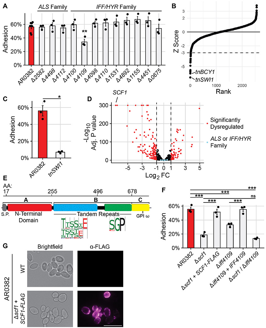
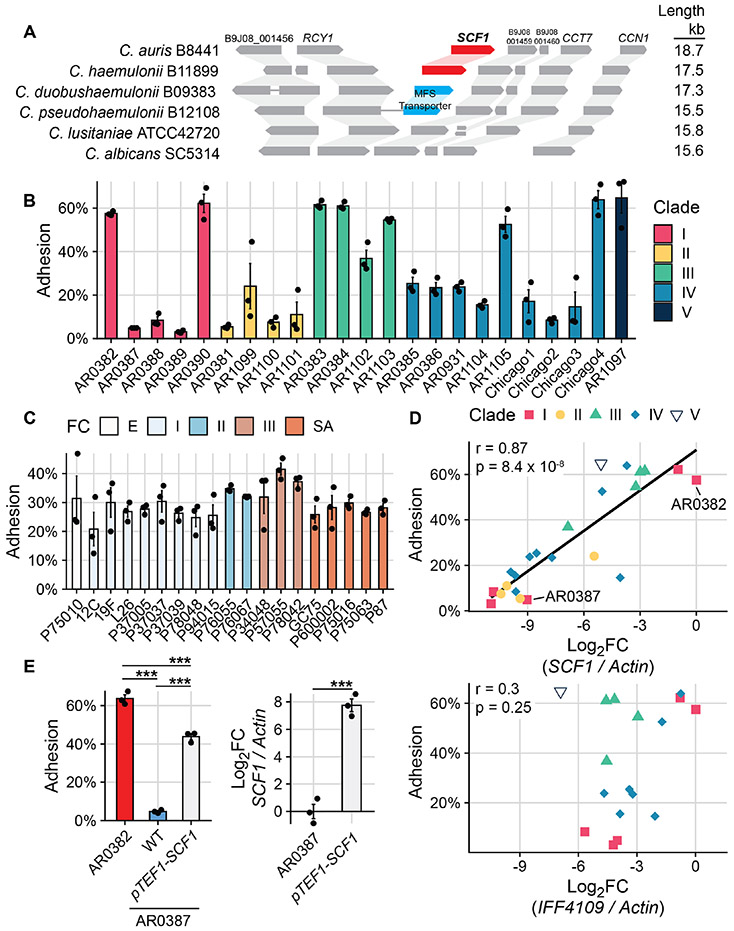
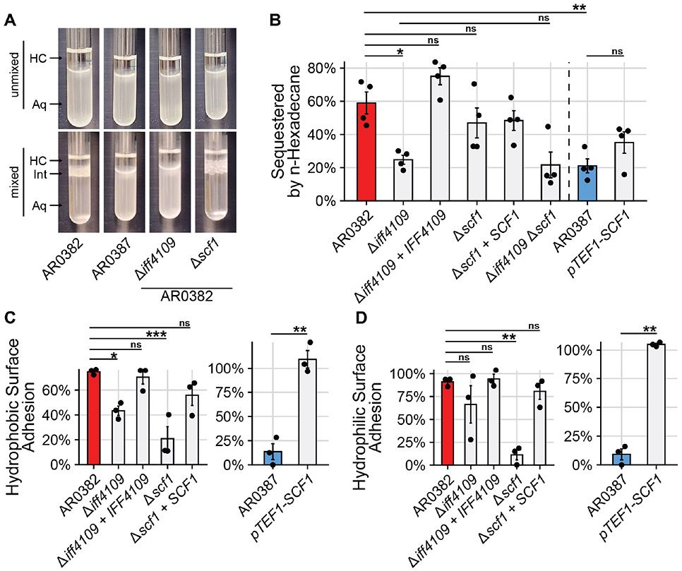
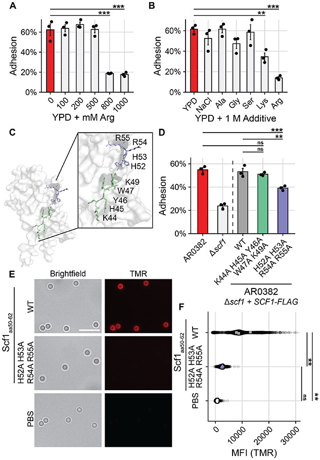
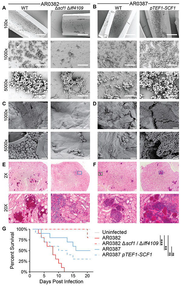
Candida auris is an emerging fungal pathogen responsible for health care-associated outbreaks that arise from persistent surface and skin colonization. We characterized the arsenal of adhesins used by C. auris and discovered an uncharacterized adhesin, Surface Colonization Factor (Scf1), and a conserved adhesin, Iff4109, that are essential for the colonization of inert surfaces and mammalian hosts. SCF1 is apparently specific to C. auris, and its expression mediates adhesion to inert and biological surfaces across isolates from all five clades. Unlike canonical fungal adhesins, which function through hydrophobic interactions, Scf1 relies on exposed cationic residues for surface association. SCF1 is required for C. auris biofilm formation, skin colonization, virulence in systemic infection, and colonization of inserted medical devices.


Figures

Fig. 1.
Fig. 1.. Surface Colonization Factor (SCF1) mediates C. auris adhesion to polymer surfaces.
(A) Adhesion of wild type AR0382 or mutants lacking one of twelve genes from ALS or IFF/HYR adhesin families. (B) 2,560 insertional mutants in the AR0382 strain background were screened for adhesion defects by measuring the proportion of cells able to remain attached to a cyclic olefin polymer surface after 3 washes with PBS. Strains are ordered by Z-score rank. Mutants with a Z-score more negative than −3 were considered to have a significant adhesive defect. (C) Adhesion of AR0382 and an insertional SWI1 mutant. (D) RNA-seq comparing the transcriptome of tnSWI1 to AR0382. SCF1 (B9J08_001458) is the strongest dysregulated gene. (E) Predicted domain architecture of Scf1, based on the clade I primary sequence, is consistent with canonical fungal adhesins. (F) Adhesion of adhesin mutants and complements compared to AR0382 (G) Immunofluorescence microscopy using an α-FLAG antibody. Representative images shown for WT AR0382 and AR0382 Δscf1 + SCF1-FLAG. Scale bar = 5 μm. Statistical differences were assessed using one-way ANOVA with Dunnett’s post-hoc test (A), student’s t-test (C), or one-way ANOVA with Tukey’s post-hoc test (G); *p ≤ 0.05; **p ≤ 0.01; ***p ≤ 0.001; ns: p > 0.05.
Fig. 2.
Fig. 2.. C. auris uniquely relies on SCF1 for adhesive plasticity.
(A) Synteny schema depicting SCF1 and the conservation and orientation of adjacent ORFs. Genomic loci are shown in comparison to C. auris. Putative SCF1 homologs were only identified in C. auris and C. haemulonii. (B) Adhesion of 23 C. auris clinical isolates from all 5 clades. (C) Adhesion of 19 C. albicans clinical isolates from five clades. FC = Fingerprint Clade. (D) SCF1 transcript abundance (top panel) but not IFF4109 transcript abundance (bottom panel) is associated with adhesion to polystyrene in the same 23 C. auris isolates from (A). Log2FC are expressed relative to AR0382. Each point signifies the mean of three biological replicates. Pearson correlation coefficient and p-value indicated. Isolates that do not encode IFF4109 are not indicated in the bottom panel. (E) Comparison of adhesion between two Clade I isolates: AR0382 and AR0387. Overexpression of SCF1 using the strong TEF1 promoter (right panel) is sufficient to drive adhesion in the poorly adhesive AR0387 background (left panel). Statistical differences were assessed using one-way ANOVA (A) and (B), with Tukey’s post-hoc test (D) or student’s t-test (D); *p ≤ 0.05; **p ≤ 0.01; ***p ≤ 0.001; ns: p > 0.05.
Fig. 3.
Fig. 3.. IFF4109, but not SCF1, mediates adhesion through cell surface hydrophobicity.
(A) Representative images from Microbial Attachment to Hydrocarbons (MATH) assay. Hydrophobic cells are sequestered from the aqueous phase (Aq) to the aqueous-hydrocarbon interface (Int) after mixing with the hydrocarbon phase (HC). (B) Proportion of cells sequestered out of the aqueous phase during MATH assay. (C), (D) Cells were allowed to attach to a hydrophobic, untreated polystyrene surface (C) or a hydrophilic, vacuum plasma treated polystyrene surface (D) for 1 hour. The surface was washed and the proportion of cells that remained attached after washing was measured. Statistical differences were assessed using one-way ANOVA with Tukey’s post-hoc test (B), (C), and (D) or student’s t-test (C) and (D); *p ≤ 0.05; **p ≤ 0.01; ***p ≤ 0.001; ns: p > 0.05.
Fig. 4.
Fig. 4.. Specific cationic residues are critical for Scf1-mediated surface association
(A), (B) WT AR0382 adhesion in the presence of increasing concentrations of arginine (A) or 1 M additives (B). (C) Predictive model of the Scf1 N-terminal domain with two neighboring cationic-aromatic clusters highlighted. (D) Adhesion of wild type AR0382, a mutant lacking SCF1, or AR0382 Δscf1 + SCF1-FLAG mutants encoding the wild type SCF1 allele or alleles containing the indicated mutations. (E) TMR-labelled 13-amino acid peptides corresponding to the wild type Scf1 sequence (residues 50-62) or the same sequence with the indicated mutations incubated with the same polystyrene microspheres used to measure adhesion in (D). Scale Bar = 5 μm. (F) Quantification (MFI) of peptide binding to individual polystyrene microspheres as in (E) measured by TMR epifluorescence, corrected for background fluorescence. Each point represents an individual microsphere. Colored points represent averages of individual experiments, used for statistical analysis. Statistical differences were assessed using one-way ANOVA with Dunnett’s post-hoc test (A), (B) or one-way ANOVA with Tukey’s post-hoc test (D) and (F); *p ≤ 0.05; **p ≤ 0.01; ***p ≤ 0.001; ns: p > 0.05.
Fig. 5.
Fig. 5.. SCF1 mediates host colonization and infection phenotypes.
(A), (B) Polyethylene central venous catheters were set in rat jugular veins and inoculated intraluminally with C. auris. Representative scanning electron microscopy images of the luminal catheter surface are shown from catheters collected 24 hrs after infection. Scale bars: 400 μm (100x), 40 μm (1000x), 5 μm (5000x). (C), (D) Full thickness human skin explants were colonized with C. auris for 24 hrs before washing to remove unassociated cells. Representative scanning electron microscopy images of the skin surface following washing are shown. Scale bars: 20 μm (1000x), 4 μm (5000x). (E), (F), (G) Immunosuppressed mice were infected intravenously (via tail vein injection) with 5 x 10⁷ C. auris cells. Histopathology sections of the kidneys 7 days post infection (E), (F) were stained with PAS. Magenta color indicates lesion areas. Ten infected mice for each strain were monitored for survival for 21 days (G). Statistical comparisons of overall survival were assessed using the Mantel-Haenszel log-rank test with Benjamini-Hochberg correction. *p ≤ 0.05; **p ≤ 0.01; ***p ≤ 0.001; ns p > 0.05.

Similar articles

Cited by

References

    1. Chakrabarti A, Sood P, On the emergence, spread and resistance of Candida auris: host, pathogen and environmental tipping points. J. Med. Microbiol 70 (2021), doi:10.1099/jmm.0.001318. - DOI - PMC - PubMed
    1. Akinbobola AB, Kean R, Hanifi SMA, Quilliam RS, Environmental reservoirs of the drug-resistant pathogenic yeast Candida auris. PLoS Pathog. 19, e1011268 (2023). - PMC - PubMed
    1. Ashkenazi-Hoffnung L, Rosenberg Danziger C, Navigating the New Reality: A Review of the Epidemiological, Clinical, and Microbiological Characteristics of Candida auris, with a Focus on Children. J Fungi (Basel). 9 (2023), doi:10.3390/jof9020176. - DOI - PMC - PubMed
    1. Chow NA, Gade L, Tsay SV, Forsberg K, Greenko JA, Southwick KL, Barrett PM, Kerins JL, Lockhart SR, Chiller TM, Litvintseva AP, US Candida auris Investigation Team, Multiple introductions and subsequent transmission of multidrug-resistant Candida auris in the USA: a molecular epidemiological survey. Lancet Infect. Dis 18, 1377–1384 (2018). - PMC - PubMed
    1. Roberts SC, Zembower TR, Ozer EA, Qi C, Genetic Evaluation of Nosocomial Candida auris Transmission. J. Clin. Microbiol 59 (2021), doi:10.1128/JCM.02252-20. - DOI - PMC - PubMed