. 2023 Dec 1;146(12):5000-5014.
doi: 10.1093/brain/awad331.

Neuromelanin accumulation drives endogenous synucleinopathy in non-human primates

Affiliations

Neuromelanin accumulation drives endogenous synucleinopathy in non-human primates

Julia Chocarro et al. Brain. .

Abstract

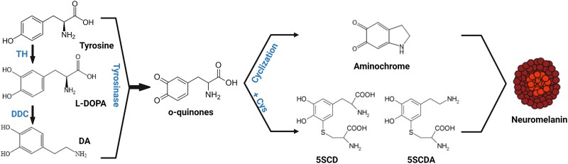
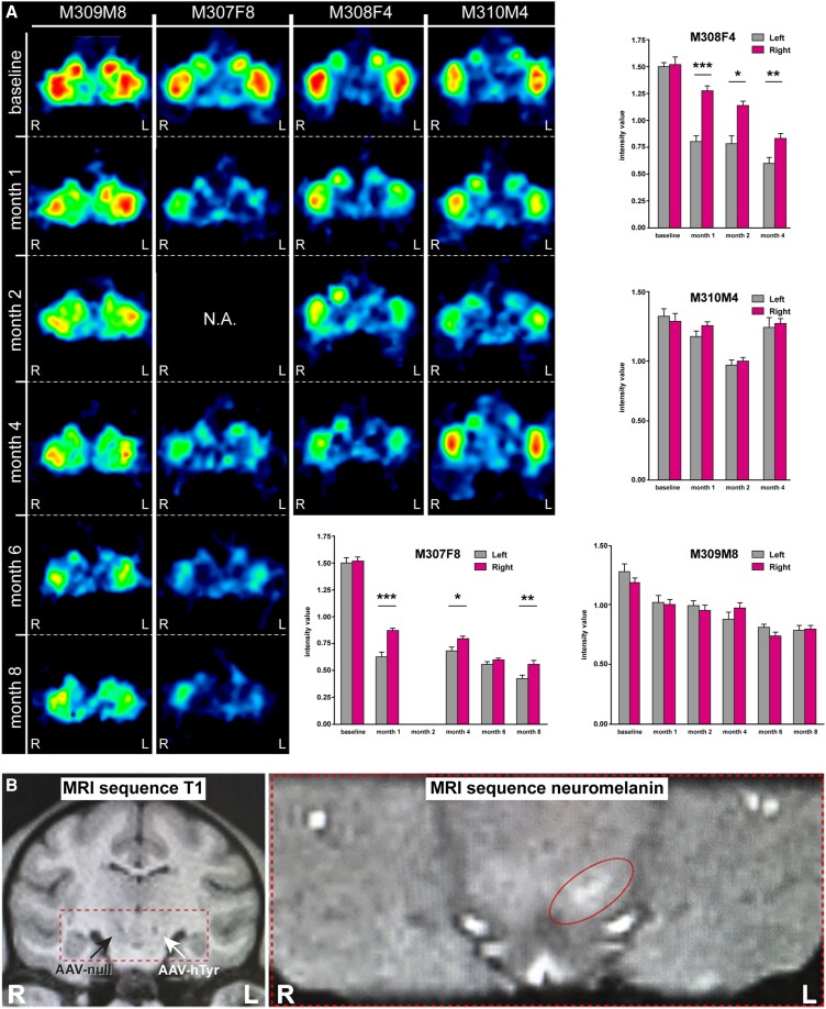
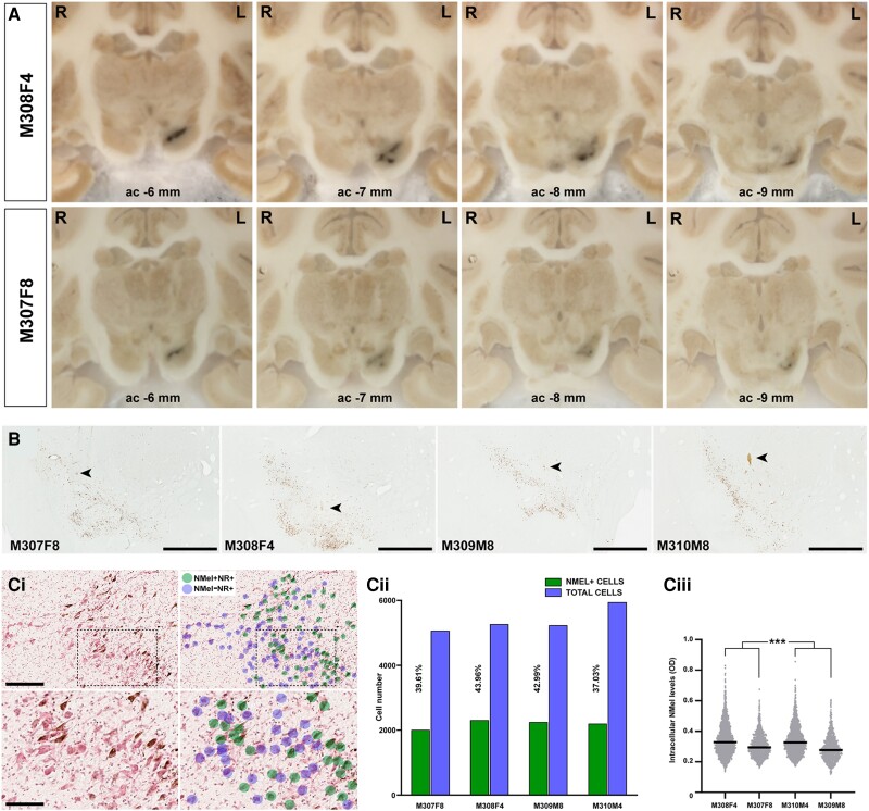
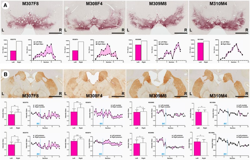
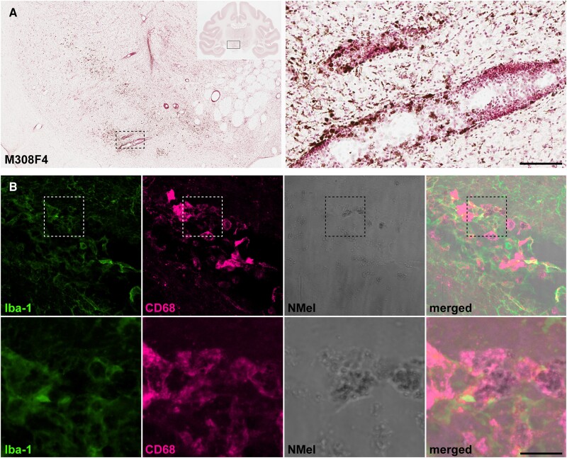
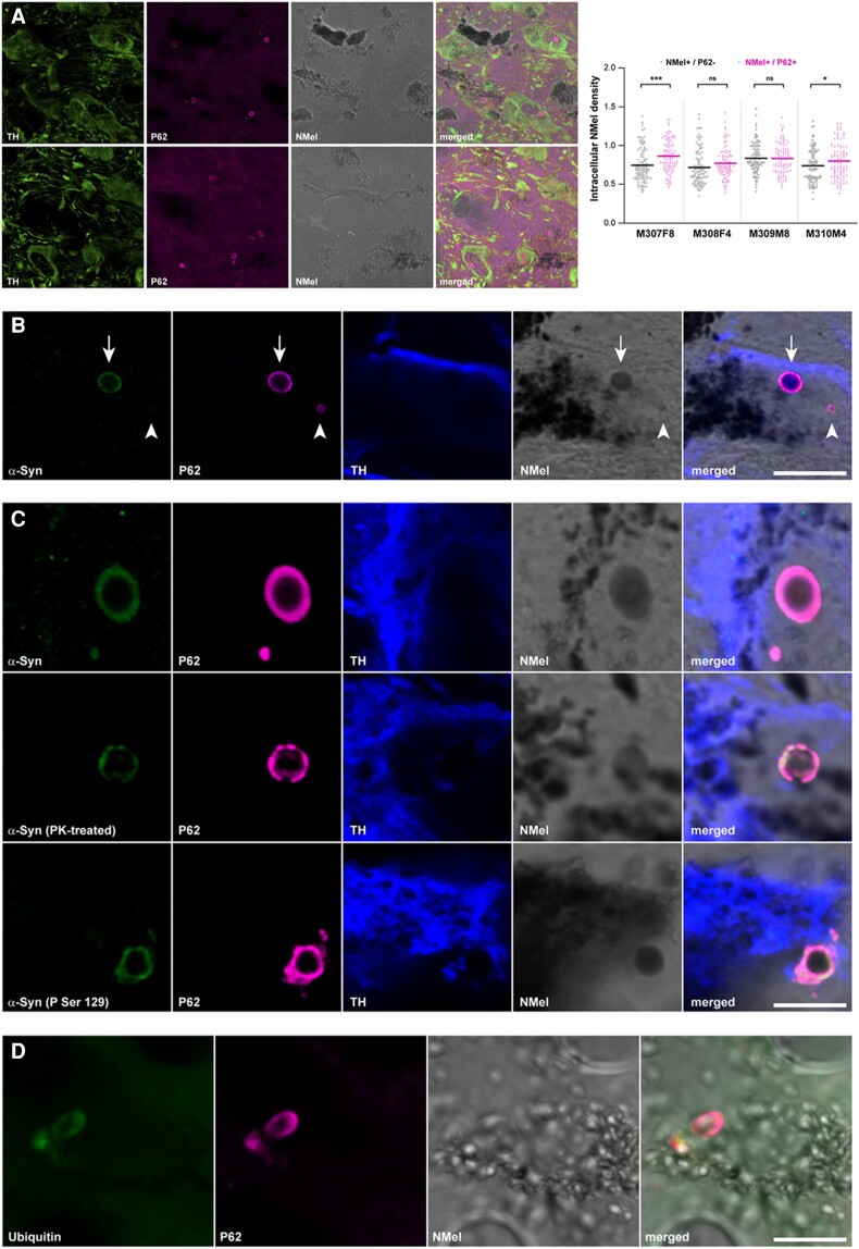
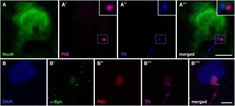
Although neuromelanin is a dark pigment characteristic of dopaminergic neurons in the human substantia nigra pars compacta, its potential role in the pathogenesis of Parkinson's disease (PD) has often been neglected since most commonly used laboratory animals lack neuromelanin. Here we took advantage of adeno-associated viral vectors encoding the human tyrosinase gene for triggering a time-dependent neuromelanin accumulation within substantia nigra pars compacta dopaminergic neurons in macaques up to similar levels of pigmentation as observed in elderly humans. Furthermore, neuromelanin accumulation induced an endogenous synucleinopathy mimicking intracellular inclusions typically observed in PD together with a progressive degeneration of neuromelanin-expressing dopaminergic neurons. Moreover, Lewy body-like intracellular inclusions were observed in cortical areas of the frontal lobe receiving dopaminergic innervation, supporting a circuit-specific anterograde spread of endogenous synucleinopathy by permissive trans-synaptic templating. In summary, the conducted strategy resulted in the development and characterization of a new macaque model of PD matching the known neuropathology of this disorder with unprecedented accuracy. Most importantly, evidence is provided showing that intracellular aggregation of endogenous α-synuclein is triggered by neuromelanin accumulation, therefore any therapeutic approach intended to decrease neuromelanin levels may provide appealing choices for the successful implementation of novel PD therapeutics.

Keywords: Lewy bodies; alpha-synuclein; marinesco bodies; prion-like spread; tyrosinase.


Conflict of interest statement

The authors report no competing interests.

Figures

Figure 1
Figure 1
Schematic representation of neuromelanin synthesis. Dopamine is synthesized in cytoplasm from L-DOPA by DDC. Both cytosolica dopamine (DAD and L-DOPA) can be oxidized either spontaneously or by tyrosinase to produce o-quinones. These, in turn generate aminochrome, 5SDCA and 5SCD, which will act as precursors of the melanic components of neuromelanin. 5SCD = 5-S-cysteinyldopa; 5SCDA = 5-5cysteinyldopamine; DDC = DOPA-decarboxylase; L-DOPA = 3,4-dihydroxyphenylalanine; TH = tyrosine hydroxylase. Figure was created by Marta González-Sepúlveda with BioRender.com (agreement No. JI25JEO0Y7).
Figure 2
Figure 2
Neuroimaging studies. (A) MicroPET studies. Representative coronal sections of the non-human primate (NHP) brain at the level of the putamen nucleus showing the binding potential of 11C-DTBZ (a selective VMAT2 ligand). Histograms illustrate the obtained measurements by comparing radiotracer uptake in left versus right hemispheres at each pre-defined time point. Statistical differences were more often found in female animals. *P < 0.05, **P < 0.01, ***P < 0.001, related samples t-test, n = 10 sections/animal. Data are presented as mean ± standard deviation (SD). (B) MRI scans. Left: Anatomical T1-weighted coronal MRI scan taken at the level of the ventral mesencephalon showing the location of the injection sites for AAV-hTyr (left SNpc) and the control AAV-null (right SNpc). Right: Hyperintense area in the left SNpc as observed with a neuromelanin-dedicated sequence. SNpc = substantia nigra pars compacta.
Figure 3
Figure 3
Pigmentation of the substantia nigra upon the delivery of AAV-hTyr. (A) Macroscopic images of non-human primate (NHP) brains taken from the microtome during sectioning. A darkened area at the level of the left substantia nigra pars compacta (SNpc) is visible to the naked eye throughout the whole rostrocaudal extent of this structure. A time-dependent loss of pigmentation is observed when comparing animals with post-viral injections follow-up times of 4 and 8 months. (B) Low-power microphotographs taken from non-stained sections of all animals at the level of the SNpc. Pigmentation with neuromelanin is clearly visible even at low magnification. Arrowheads indicate the location of the injection sites. Scale bar = 1000 μm. [C(i)] Illustrative example showing the conducted procedure for quantifying the number of neurons being transduced with AAV-hTyr in the SNpc. By taking advantage of sections counterstained with neutral red (NR), a bi-layered algorithm was prepared with Aiforia® (www.aiforia.com) to further disclose pigmented and non-pigmented NR+ neurons in the SNpc. [C(ii)] Histogram showing the percentages of pigmented neurons in the SNpc resulting from the use of the Aiforia® algorithm. [C(iii)] Box plot showing the distribution and mean values of intracellular neuromelanin levels in all animals. neuromelanin densities in female animals are higher than in males at each time point post-injection of AAV-hTyr. ***P < 0.001, nested ANOVA test with time and gender as fixed factors, and monkeys nested within fixed factors. Measurements were conducted in 12 consecutive sections covering the entire rostrocaudal extent of the SNpc (equally spaced 400 μm each). AAV = adeno-associated viral vectors; hTyr = human tyrosinase gene.
Figure 4
Figure 4
Time-dependent dopaminergic nigrostriatal damage driven by neuromelanin accumulation. (A) Coronal sections taken at the level of the substantia nigra pars compacta (SNpc) immunostained with an antibody against tyrosine hydroxylase (TH) and visualized with a purple peroxidase chromogen (V-VIP). Histograms below illustrate the number of TH+ cells in the left versus right SNpc, together with the rostrocaudal distribution of cell numbers in the SNpc (www.aiforia.com). Scale bar = 3200 μm. (B) Coronal sections taken at the level of the post-commissural caudate and putamen nuclei immunostained for TH and visualized with a brown peroxidase chromogen (DAB). Scale bar = 6400 μm. Histograms below illustrate optical densities of TH in the left and right caudate and putamen nuclei for each animal, together with the rostrocaudal distribution. Dashed blue lines labelled as ‘ac’ indicate the position of the anterior commissure. **P < 0.01, ***P < 0.001, unpaired t-test, n = 20 sections/animal. Data are presented as mean ± SD.
Figure 5
Figure 5
Extracellular neuromelanin accumulation and pro-inflammatory scenario. (A) Coronal section through the substantia nigra pars compacta (SNpc) in Animal M308F4 counterstained with neutral red and showing the distribution of extracellular neuromelanin resulting from the loss of dopaminergic cells. Inset illustrates the preferential perivascular location of extracellular neuromelanin deposits. Scale bar = 1000 μm (left) and 1000 μm (inset). (B) Confocal images showing the pro-inflammatory scenario made by cells positive for Iba-1 (green channel) and CD68 (purple channel) that are digesting extracellular neuromelanin. Scale bar = 64 μm (AD) and 18 μm (A’D’).
Figure 6
Figure 6
Intracellular inclusions in pigmented dopaminergic neurons of the substantia nigra. (A) Confocal images showing the relative abundance of intracellular inclusions within pigmented dopaminergic neurons in the substantia nigra pars compacta (SNpc). TH+ neurons are illustrated in the green channel and P62+ inclusions in the purple channel. Neuromelanin (Nmel) is visualized under brightfield illumination. Images are taken from Animal M310M4 (top) and Anomal M307F8 (bottom). Scale bar = 100 μm. The box plot illustrates neuromelanin levels in pigmented neurons with and without P62+ intracellular inclusions. Analyses were conducted in 12 consecutive sections covering the entire rostrocaudal extent of the SNpc (equally spaced 400 μm each). (B) Two different types of intracellular inclusions were observed in pigmented dopaminergic neurons (TH+; blue channel), namely Marinesco bodies (MBs, intranuclear, arrowhead) and Lewy bodies (LBs, intracytoplasmic, arrow). While Lewy bodies are positive for both P62 (purple channel) and α-Syn (green channel), Marinesco bodies only displayed immunoreactivity for P62. Images are taken from Animal M308F4. Scale bar = 15 μm. (C) Lewy body-like intracytoplasmic inclusions within pigmented dopaminergic cells (TH+, blue channel) are made of insoluble and phosphorylated α-Syn species (green channel). Observed P62+ inclusions (purple channel) are also labelled with antibodies against either total or phosphorylated α-Syn (top and bottom, respectively) and are resistant to digestion with proteinase K (middle). All images are taken from Animal M307F8. Scale bar = 5 μm. (D) Intracytoplasmic inclusions (e.g. Lewy body-like) observed in neuromelanized neurons are positive for P62 (purple channel) as well as for ubiquitin (green channel). Scale bar = 15 μm. TH = tyrosine hydroxylase.
Figure 7
Figure 7
Anterograde spread of endogenous α-synuclein towards the prefrontal cerebral cortex. (AA’’’) Confocal images taken at the level of the superior frontal gyrus in Animal M309M8 showing a layer V pyramidal neuron (stained with NeuN; green channel) receiving a TH+ terminal (blue channel) and showing an intracytoplasmic inclusion (positive for P62) just opposite to the arrival of the terminal bouton. Scale bar = 10 μm. (BB’’’) Confocal images taken from the anterior cingulate gyrus in Animal M307F8 showing that P62+ intracytoplasmic inclusions (red channel) are also positive for α-Syn (green channel) and are observed in layer V neurons receiving TH+ terminals (purple channel). Scale bar = 20 μm. TH = tyrosine hydroxylase.

Comment in

References

    1. Fedorow H, Tribl F, Halliday G, Gerlanch M, Riederer P, Double KL. Neuromelanin in human dopamine neurons: Comparison with peripheral melanins and relevance to Parkinson’s disease. Prog Neurobiol. 2005;75:109–124. - PubMed
    1. Fedorow H, Halliday GM, Rickert CH, Gerlach M, Riederer P, Double KL. Evidence for specific phases in the development of human neuromelanin. Neurobiol Aging 2006;27:506–512. - PubMed
    1. Hirsch EC, Graybiel AM, Agid Y. Melanized dopaminergic neurons are differentially susceptible to degeneration in Parkinson’s disease. Nature 1988;334:345–348. - PubMed
    1. Hirsch EC, Graybiel AM, Agid Y. Selective vulnerability of pigmented dopaminergic neurons in Parkinson’s disease. Acta Neurol Scand. 1989;126:19–22. - PubMed
    1. Kastner A, Hirsch EC, Lejeune O, Javoy-Agid F, Rascol O, Agid Y. Is the vulnerability of neurons in the substantia nigra of patients with Parkinson’s disease related to their neuromelanin content? J Neurochem. 1992;59:1080–1089. - PubMed

Publication types