. 2023 Oct 15:336:199231.
doi: 10.1016/j.virusres.2023.199231. Epub 2023 Sep 28.

Biophysical characterization of the cetacean morbillivirus haemagglutinin glycoprotein

Affiliations

Biophysical characterization of the cetacean morbillivirus haemagglutinin glycoprotein

Luca Zinzula et al. Virus Res. .

Abstract

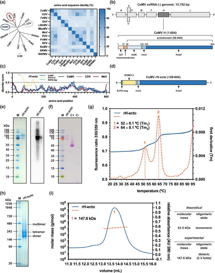
Cetacean morbillivirus (CeMV) is an enveloped, non-segmented, negative-stranded RNA virus that infects marine mammals, spreading across species and causing lethal disease outbreaks worldwide. Among the eight proteins encoded by the CeMV genome, the haemagglutinin (H) glycoprotein is responsible for the virus attachment to host cell receptors. CeMV H represents an attractive target for antiviral and diagnostic research, yet the elucidation of the molecular mechanisms underlying its role in infection and inter-species transmission was hampered thus far due to the unavailability of recombinant versions of the protein. Here we present the cloning, expression and purification of a recombinant CeMV H ectodomain (rH-ecto), providing an initial characterization of its biophysical and structural properties. Sodium dodecyl sulphate - polyacrylamide gel electrophoresis (PAGE) combined to Western blot analysis and periodic acid Schiff assay showed that CeMV rH-ecto is purifiable at homogeneity from insect cells as a secreted, soluble and glycosylated protein. Miniaturized differential scanning fluorimetry, Blue Native PAGE and size exclusion chromatography coupled to multiangle light scattering revealed that CeMV rH-ecto is globularly folded, thermally stable and exists in solution in the oligomeric states of dimer and multiple of dimers. Furthermore, negative stain electron microscopy single particle analysis allowed us to delineate a low-resolution molecular architecture of the CeMV rH-ecto dimer, which recapitulates native assemblies from other morbilliviral H proteins, such as those from measles virus and canine distemper virus. This set of experiments by orthogonal techniques validates the CeMV rH-ecto as an experimental model for future biochemical studies on its structure and functions.

Keywords: Cetacean morbillivirus; Cetaceans; Haemagglutinin; Host–pathogen interaction; Morbilliviruses; Viral pathogenesis.


Conflict of interest statement

Declaration of Competing Interest The authors declare that they have no known competing financial interests or personal relationships that could have appeared to influence the work reported in the paper.

Figures

Fig 1
Fig. 1
Design and biophysical characterization of CeMV rH-ecto. (a) Molecular evolutionary analysis of the H proteins from members of the Morbillivirus genus based on a maximum-likelihood phylogenetic tree and an amino acid sequence identity heatmap, both built from a MSA of the NCBI Protein RefSeq GenBank IDs: CDV, NP_047206.1; CeMV, NP_945029.1; FeMV, YP_009512963.1; GaV, UQM99546.1; LBbMV-1, UBB42344.1; MbMV, UBB97712.1; MeV, NP_056923.1; PDV, YP_009177603.1; PoMV, QWQ56142.1; RPV, YP_087125.2; RoMV, DAZ91188.1, SRMV, YP_133827.2; WNfMV-1, UBB42351.1; the target of this work and orthologs with available protein structure are highlighted as circled in regal-blue (CeMV), bright-red (MeV) and lilac (CDV) color in the tree. (b) CeMV genome organization (upper panel), with H gene highlighted in dark gray, and structural layout of encoded H protein (lower panel) with tail (champagne) and transmembrane (tacao) domain and stalk (link-water), neck (cornflower) and head (astral) subdomains highlighted in color. (c) IUPre3 comparative prediction of intrinsic disorder and structural domain organization profile between CeMV (regal-blue), CeMV rH-ecto (downy, cream-brulee, astral), CDV (lilac) and MeV (bright red) H proteins. (d) schematic representation of the structural organization of pCoofy64-SUMO3-CeMV-rH-ecto construct; His6-tag and SUMO-3 fusion protein are represented as cylinders colored in downy and cream-brulee, respectively. (e) Blue Coomassie-stained SDS-PAGE (left), and anti-His6 WB analysis (right) of purified CeMV rH-ecto (M, molecular weight marker). (f) WB coupled to PAS glycosylation assay of CeMV rH-ecto; magenta-colored bands are indicative of the Schiff bonds presence and of the protein glycosylated state (M, molecular weight marker; C+ and C-, positive and negative controls, respectively). (g) Nano-DSF analysis of purified CeMV rH-ecto; thermal stability and conformational profile is shown by superimposed F330/350 ratio (azure) and first derivative (outrageous-orange); conformational transitions corresponding to inflections in intrinsic fluorescence and related Tm1 (52.0 ± 0.1 °C) and Tm2 (64.0 ± 0.1 °C) are indicated as dashed and dotted lines, respectively. Data points represent averages of three independent experiments. (h) BN-PAGE analysis of purified CeMV rH-ecto (M, molecular weight marker). (i) SEC-MALS analysis (left panel) of purified CeMV rH-ecto; relative absorbance (azure) and absolute molecular mass (outrageous-orange) are indicated as continuous and dashed colored lines, respectively; the main peak (1) at which the average absolute mass (147.6 kDa) and its stoichiometry with respect to expected monomeric mass (2.3 folds) were calculated is indicated; a table (right panel) summarizing the theoretically expected monomeric mass and the experimentally determined oligomeric one is shown.
Fig 2
Fig. 2
Low-resolution molecular architecture of CeMV rH-ecto. (a) negative stain EM micrograph of purified CeMV rH-ecto (62,000 ×; scale bar, 50 nm), with three representative single particles in the zoomed insets. (b) selected 2D class averages (left panel) of dimeric CeMV rH-ecto particles, and 3D volume reconstruction (right panel) of dimeric CeMV rH-ecto, shown in three orientations. (c) comparative volumetric analysis (upper panel) of 3D-reconstructed dimeric CeMV rH-ecto with the volumes of dimeric MeV and CDV H ectodomains obtained by downfiltering to 20 Å the respective crystal structures. Atomic model (lower panel) of dimeric CeMV rH-ecto modelled with ColabFold AlphaFold2 and fitted into the SPA 3D-reconstructed volume, shown in two orientations; its spatial arrangement with respect to the remaining portions of the H proteins and the viral envelope is shown in the cartoon. (d) Schematic view of the 3D-reconstructed volume and fitted atomic model of dimeric CeMV rH-ecto (regal-blue) complexed to SLAM (reef-green) and nectin-4 (chestnut-rose), shown in two orientations, obtained by structural alignment with corresponding crystallographic structures of MeV in complex with the two receptors; SLAM and nectin-4 are purposedly shown simultaneously to highlight partial overlap of their putative binding sites.

Similar articles

References

    1. Baker J.D., Harting A.L., Barbieri M.M., Robinson S.J., Gulland F.M.D., Littnan C.L. Modeling a morbillivirus outbreak in hawaiian monk seals (Neomonachus schauinslandi) to aid in the design of mitigation programs. J. Wildl. Dis. 2017;53:736–748. doi: 10.7589/2016-10-238. - DOI - PubMed
    1. Banyard A.C., Tiwari A., Barrett T. Morbillivirus infection in pilot whales: strict protein requirement drives genetic conservation. Arch. Virol. 2011;156:1853–1859. doi: 10.1007/s00705-011-1042-8. - DOI - PubMed
    1. Beffagna G., Centelleghe C., Franzo G., Di Guardo G., Mazzariol S. Genomic and structural investigation on dolphin morbillivirus (DMV) in Mediterranean fin whales (Balaenoptera physalus) Sci. Rep. 2017;7:41554. doi: 10.1038/srep41554. - DOI - PMC - PubMed
    1. Bellière E.N., Esperón F., Fernández A., Arbelo M., Muñoz M.J., Sánchez-Vizcaíno J.M. Phylogenetic analysis of a new Cetacean morbillivirus from a short-finned pilot whale stranded in the Canary Islands. Res. Vet. Sci. 2011;90:324–328. doi: 10.1016/j.rvsc.2010.05.038. - DOI - PubMed
    1. Blixenkrone-Møller M., Bolt G., Jensen T.D., Harder T., Svansson V. Comparative analysis of the attachment protein gene (H) of dolphin morbillivirus. Virus Res. 1996;40:47–55. doi: 10.1016/0168-1702(95)01254-0. - DOI - PubMed

LinkOut - more resources