. 2023 Oct 15:748:109769.
doi: 10.1016/j.abb.2023.109769. Epub 2023 Sep 27.

Effects of spike proteins on angiotensin converting enzyme 2 (ACE2)

Affiliations

Effects of spike proteins on angiotensin converting enzyme 2 (ACE2)

Jennyfer Bejoy et al. Arch Biochem Biophys. .

Abstract

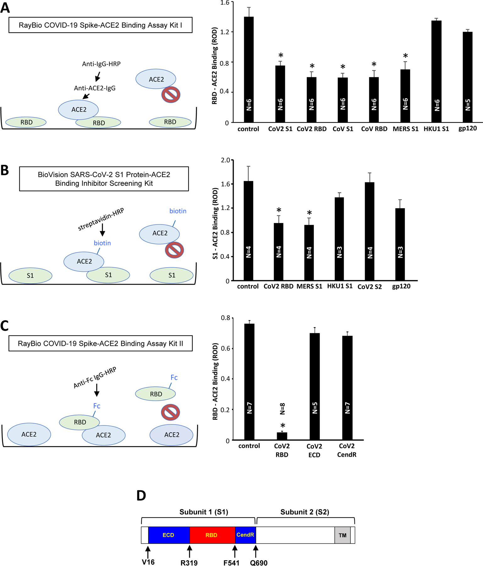
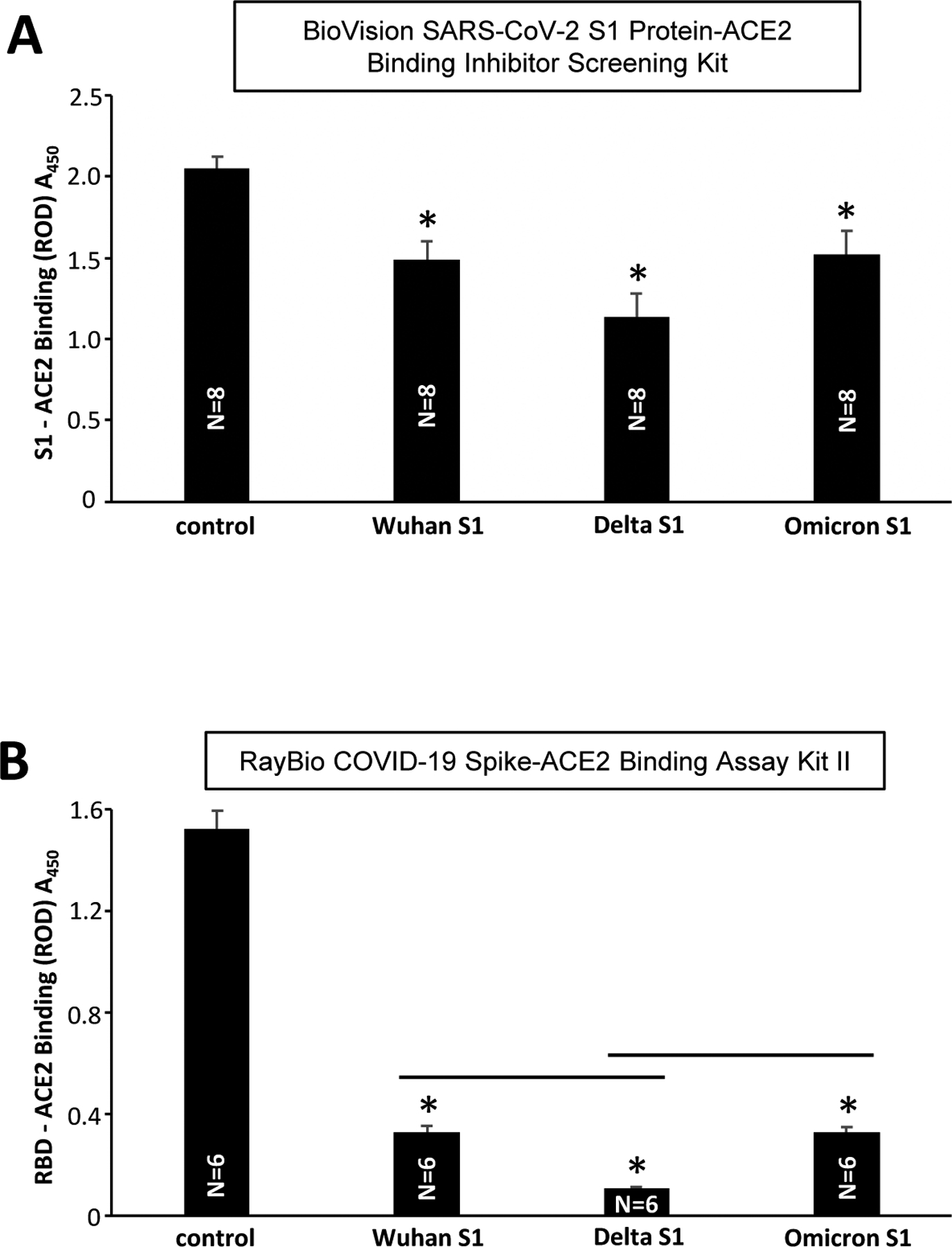
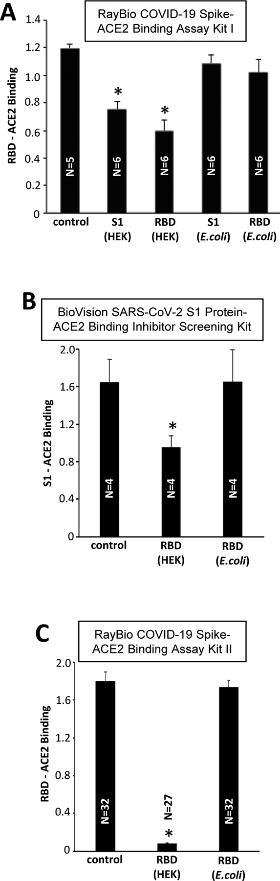
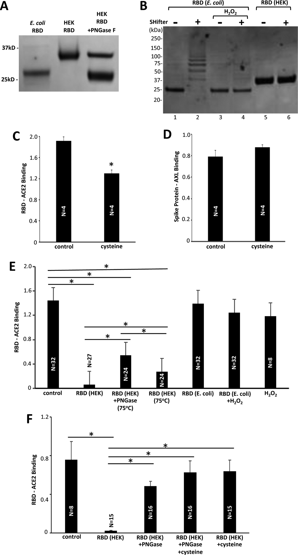
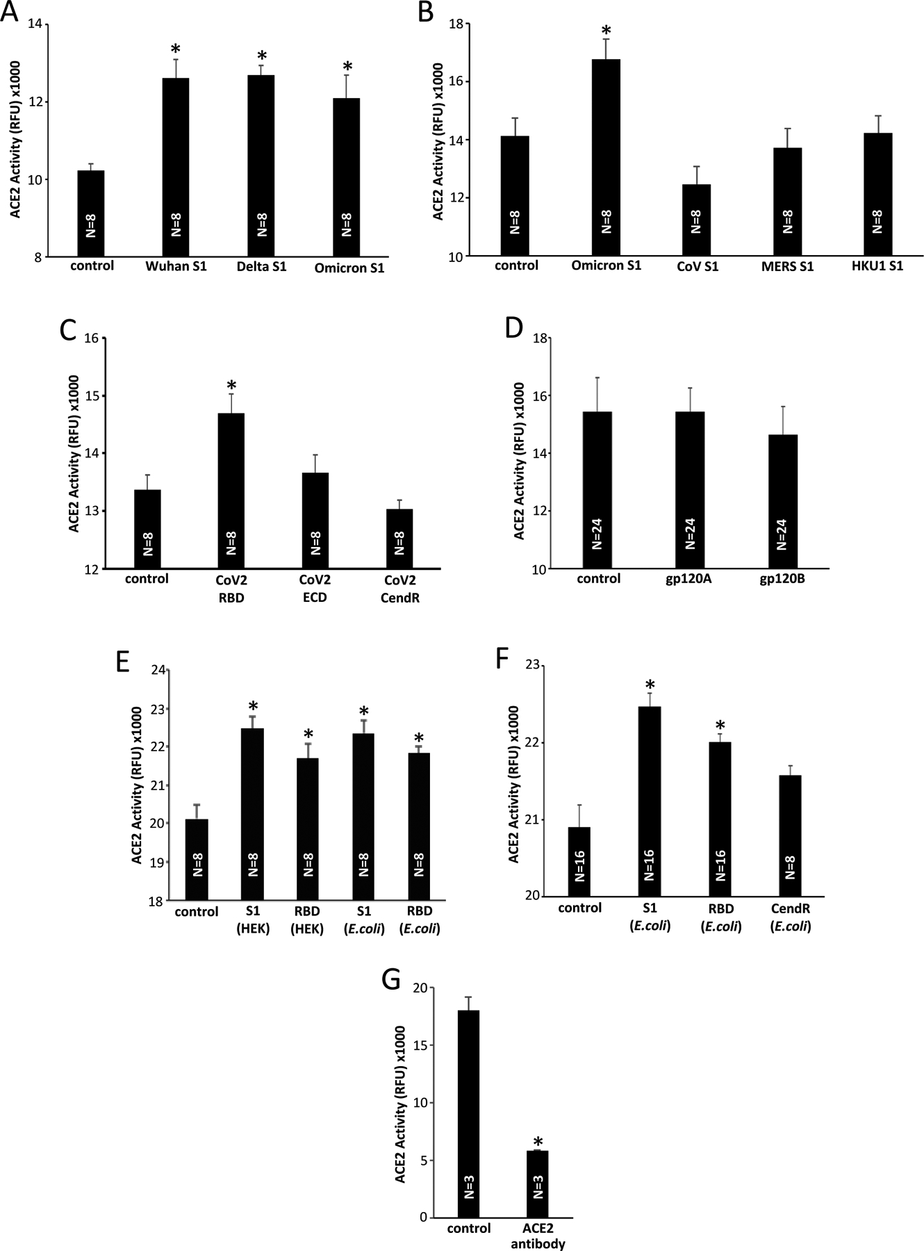
The Coronavirus Disease 2019 (COVID-19) pandemic was caused by Severe Acute Respiratory Syndrome Coronavirus 2 (SARS-CoV-2), which enters host cells through interactions of its spike protein to Angiotensin-Converting Enzyme 2 (ACE2). ACE2 is a peptidase that cleaves Angiotensin II, a critical pathological mediator. This study investigated if the spike protein binding to ACE2 compromises its peptidase activity. Spike/ACE2 Binding Assays suggested that spike proteins of SARS-CoV-2, SARS-CoV and MERS-CoV, but not HKU1, bind to ACE2. S1 and receptor-binding domain (RBD), but not S2, extracellular domain (ECD) or CendR domain, bind to ACE2. While glycosylated spike proteins prepared in HEK293 cells bind to ACE2, non-glycosylated proteins produced in E. coli do not. Cysteine residues of the spike protein expressed in HEK293 cells are fully oxidized, while those of the protein expressed in E. coli are reduced. The deglycosylation of HEK cell-produced protein attenuates the ACE2 binding, while the oxidation of the E. coli protein does not promote the binding. The S1 protein of SARS-CoV-2 enhances the ACE2 peptidase activity, while SARS-CoV, MERS-CoV or HKU1 does not. The ACE2 activity is enhanced by RBD, but not ECD or CendR. In contrast to distinct ACE2 binding capacities of proteins expressed in HEK293 cells and in E. coli, spike proteins expressed in both systems enhance the ACE2 activity. Thus, the spike protein of SARS-CoV-2, but not other coronaviruses, enhances the ACE2 peptidase activity through its RBD in a glycosylation-independent manner.

Keywords: ACE2; COVID-19; Coronavirus; Peptidase; SARS-CoV-2; Spike protein.


Figures

Figure 1:
Figure 1:. The use of Spike/ACE2 Binding Assays to study the effects of spike proteins.
(A) The RayBio COVID-19 Spike-ACE2 Binding Assay Kit I has the SARS-CoV-2 RBD protein bound to the bottom of the microplate wells. The ACE2 protein is then added, washed, and bound ACE2 to RBD is detected using anti-ACE2-IgG and HRP-linked secondary antibody. The bar graph shows that the inclusion of SARS-CoV-2 spike protein S1 subunit (CoV2 S1), SARS-CoV-2 spike protein RBD-only containing protein (CoV2 RBD), SARS-CoV spike protein S1 subunit (CoV S1), SARS-CoV spike protein RBD-only containing protein (CoV RBD), and MERS-CoV spike protein S1 subunit (MERS S1) inhibited the binding of ACE2 to RBD in the kit. On the other hand, the HKU1 spike protein S1 subunit (HKU1 S1) and HIV-1 subunit B gp120 had no effects. (B) The BioVision SARS-CoV-2 S1 Protein-ACE2 Binding Inhibitor Screening Kit has the SARS-CoV-2 S1 protein bound to the bottom of the microplate wells. The biotin-linked ACE2 protein is then added, washed, and bound biotin-ACE2 to S1 is detected using streptavidin-HRP. The bar graph shows that the results of the above RayBio kit experiments were repeatable with the BioVision kit. (C) The RayBio COVID-19 Spike-ACE2 Binding Assay Kit II has the ACE2 protein bound to the bottom of the microplate wells. Fc-tagged RBD protein is then added, washed, and bound Fc-RBD to ACE2 is detected using HRP-linked anti-Fc-IgG. The bar graph shows that, while the inclusion of the SARS-CoV-2 spike protein RBD-only containing protein inhibited the binding of Fc-RBD to ACE2 in the assay kit, the ECD- or CendR domain-only containing proteins had no effects. The symbol * denotes significant difference from untreated control at P<0.05. (D) The structure of the SARS-CoV-2 spike protein. The spike protein consists of Subunit 1 (S1) and Subunit 2 (S2). The S1 subunit contains the RBD that binds to ACE2 of the host cell membrane. The S2 subunit is responsible for fusion. In this study, we used recombinant full-length S1 (Val16-Gln690), the RBD only-containing protein (Arg319-Phe541), ECD (Val16-Phe318), and CendR domain (Asn542-Arg685) (GenBank Accession Number: QHD43416.1). ROD: relative optical density. The symbol * denotes significant difference from untreated control at P<0.05.
Figure 2:
Figure 2:. Effects of SARS-CoV-2 variants on Spike/ACE2 binding.
(A) The BioVision SARS-CoV-2 S1 Protein-ACE2 Binding Inhibitor Screening Kit and (B) RayBio COVID-19 Spike-ACE2 Binding Assay Kit II were used to determine the effects of the SARS-CoV-2 spike protein S1 subunit of the original Wuhan sequence (Wu et al., 2020), the S1 subunit of the SARS-CoV-2 spike protein Delta variant, and the S1 subunit of the SARS-CoV-2 spike protein Omicron variant. The symbol * denotes significant difference from untreated control at P<0.05. The lines above bars indicates significant difference from each other at P<0.05.
Figure 3:
Figure 3:. Effects of HEK293 cell- and E. coli-expressed S1 and RBD on Spike/ACE2 binding.
The recombinant SARS-CoV-2 S1 and RBD proteins produced in HEK293 cells and E. coli were added to RBD/ACE2 Binding Assays. All the binding assay kits showed that HEK293 cell-expressed spike proteins effectively competed with the spike protein binding to ACE2 in the assay kits. By contrast, the E. coli produced proteins had no effects. * denotes significant difference from untreated control at P<0.05.
Figure 4:
Figure 4:. Mechanisms of differential actions of HEK293 cell- and E. coli-produced spike proteins.
(A) The RBD protein expressed in HEK293 cells, but not that expressed in E. coli, is glycosylated. The SARS-CoV-2 RBD-only proteins were subjected to SDS-PAGE. While the RBD protein produced in E. coli migrates at expected 25 kD, the HEK293 cell-produced RBD migrates at a higher molecular weight. The PNGase F treatment caused the HEK293 cell-expressed RBD to migrate at ~25 kD. (B) The RBD protein expressed in E. coli is reduced, but that expressed in HEK293 cells is oxidized. The SulfoBiotics Protein Redox State Monitoring assay puts a 15-kD SHifter to reduce cysteine residues resulting in electrophoretic mobility shifts. Using this assay, the RBD protein that was produced in E. coli was found to be largely reduced as the 25-kD protein (Lane 1) became shifted in the presence of the SHifter (Lane 2). On the other hand, the glycosylated RBD protein produced in HEK293 cells was found to be fully oxidized (Lane 6). H2O2 treatment fully oxidized the E. coli-produced RBD protein (Lane 4). (C) The Fc-tagged RBD protein of RayBio COVID-19 Spike-ACE2 Binding Assay Kit II was pre-treated with cysteine. * denotes significant difference from untreated control at P<0.05. (D) The S1 protein of RayBio COVID-19 Spike-AXL Binding Assay Kit was pre-treated with cysteine. (E) The SARS-CoV-2 RBD protein expressed in HEK293 cells, RBD protein expressed in HEK293 cells treated with PNGase F at 75°C, RBD protein expressed in HEK293 cells heated at 75°C, RBD protein expressed in E. coli, RBD protein expressed in E. coli treated with H2O2, and H2O2 were added to the RayBio COVID-19 Spike-ACE2 Binding Assay Kit II. (F) Reduced cysteine was also added to some of experiments shown in Panel E. Lines above bars with * indicate significant difference from each other at P<0.05.
Figure 5:
Figure 5:. Effects of spike proteins on ACE2 enzymatic activity.
(A) Various variants of SARS-CoV-2 S1 protein (40 nM) slightly but significantly enhance the ACE2 activity. (B) The S1 proteins of SARS-CoV, MERS-CoV, and HKU1 (40 nM) do not enhance the ACE2 activity. (C) Only the SARS-CoV-2 RBD protein, but not the ECD- or CendR-only protein, enhance the ACE2 activity. (D) Neither gp120 of HIV-1 subtype A (gp120A) nor subtype B (gp120B) enhance the ACE2 activity. (E) The ACE2 activity was monitored using the ACE2 Activity Assay in the presence of recombinant S1 and RBD proteins. The HEK293 cell- and E. coli-expressed proteins were equally effective in enhancing the ACE2 activity. (F) The CendR domain protein produced in E. coli does not enhance the ACE2 activity. (G) The ACE2 antibody (mouse monoclonal IgG2A; Cat# MAB9331, R&D Systems Inc., Minneapolis, MN, USA) inhibited the ACE2 activity. RFU: relative fluorescence unit. The symbol * denotes significant difference from the untreated control values at P<0.05.
Figure 6:
Figure 6:. Effects of SAR-CoV-2 spike protein S1 on other enzymes.
(A) Effects of the HEK293 cell-produced Wuhan SARS-CoV-2 spike protein S1 on the furin enzymatic activity using the Rh110 Furin Activity Assay. (B) The GAPDH activity was monitored at 450 nm in the presence of Wuhan SARS-CoV-2 spike protein S1 protein produced in HEK293 cells. (C) The Wuhan SARS-CoV-2 spike protein S1 does not influence the DPP4 activity. RFU: relative fluorescence unit. ROD: relative optical density. The symbol * denotes significant difference from the untreated control values at P<0.05.

Similar articles

Cited by

References

    1. Wu F, Zhao S, Yu B, Chen YM, Wang W, Song ZG, Hu Y, Tao ZW, Tian JH, Pei YY, Yuan ML, Zhang YL, Dai FH, Liu Y, Wang QM, Zheng JJ, Xu L, Holmes EC, Zhang YZ, A new coronavirus associated with human respiratory disease in China, Nature 579 (2020) 265–269. - PMC - PubMed
    1. Huang C, Wang Y, Li X, Ren L, Zhao J, Hu Y, Zhang L, Fan G, Xu J, Gu X, Cheng Z, Yu T, Xia J, Wei Y, Wu W, Xie X, Yin W, Li H, Liu M, Xiao Y, Gao H, Guo L, Xie J, Wang G, Jiang R, Gao Z, Jin Q, Wang J, Cao B, Clinical features of patients infected with 2019 novel coronavirus in Wuhan, China, Lancet 395 (2020) 497–506. - PMC - PubMed
    1. Petrosillo N, Viceconte G, Ergonul O, Ippolito G, Petersen E, COVID-19, SARS and MERS: are they closely related? Clin. Microbiol. Infect 26 (2020) 729–734. - PMC - PubMed
    1. Li W, Moore MJ, Vasilieva N, Sui J, Wong SK, Berne MA, Somasundaran M, Sullivan JL, Luzuriaga K, Greenough TC, Choe H, Farzan M, Angiotensin-converting enzyme 2 is a functional receptor for the SARS coronavirus, Nature 426 (2003) 450–454. - PMC - PubMed
    1. Yan R, Zhang Y, Li Y, Xia L, Guo Y, Zhou Q, Structural basis for the recognition of SARS-CoV-2 by full-length human ACE, Science 367 (2020) 1444–1448. - PMC - PubMed

Publication types

Substances

LinkOut - more resources