. 2023 Sep 28;21(1):264.
doi: 10.1186/s12964-023-01274-2.

RU.521 mitigates subarachnoid hemorrhage-induced brain injury via regulating microglial polarization and neuroinflammation mediated by the cGAS/STING/NF-κB pathway

Affiliations

RU.521 mitigates subarachnoid hemorrhage-induced brain injury via regulating microglial polarization and neuroinflammation mediated by the cGAS/STING/NF-κB pathway

Jiang Shao et al. Cell Commun Signal. .

Erratum in

Abstract

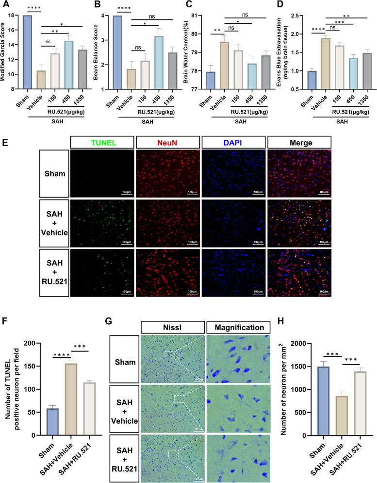
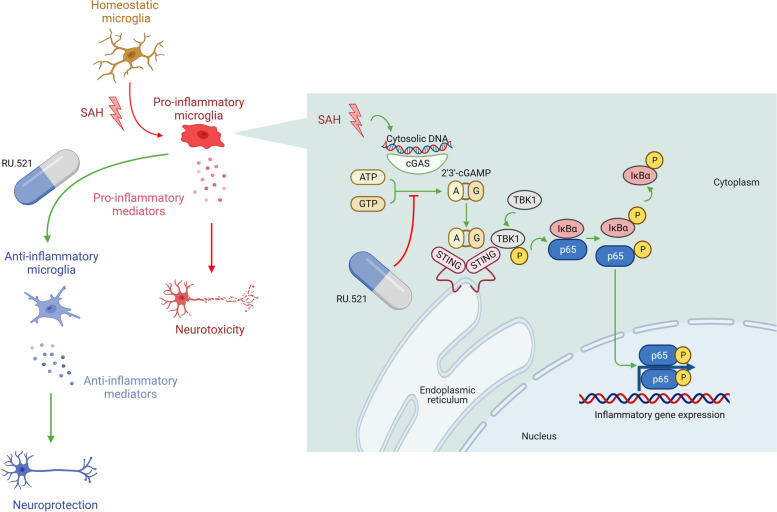
Background: The poor prognosis of subarachnoid hemorrhage (SAH) is often attributed to neuroinflammation. The cGAS-STING axis, a cytoplasmic pathway responsible for detecting dsDNA, plays a significant role in mediating neuroinflammation in neurological diseases. However, the effects of inhibiting cGAS with the selective small molecule inhibitor RU.521 on brain injury and the underlying mechanisms after SAH are still unclear.

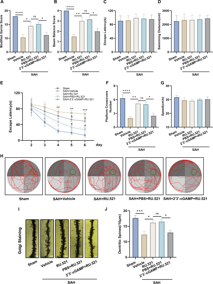
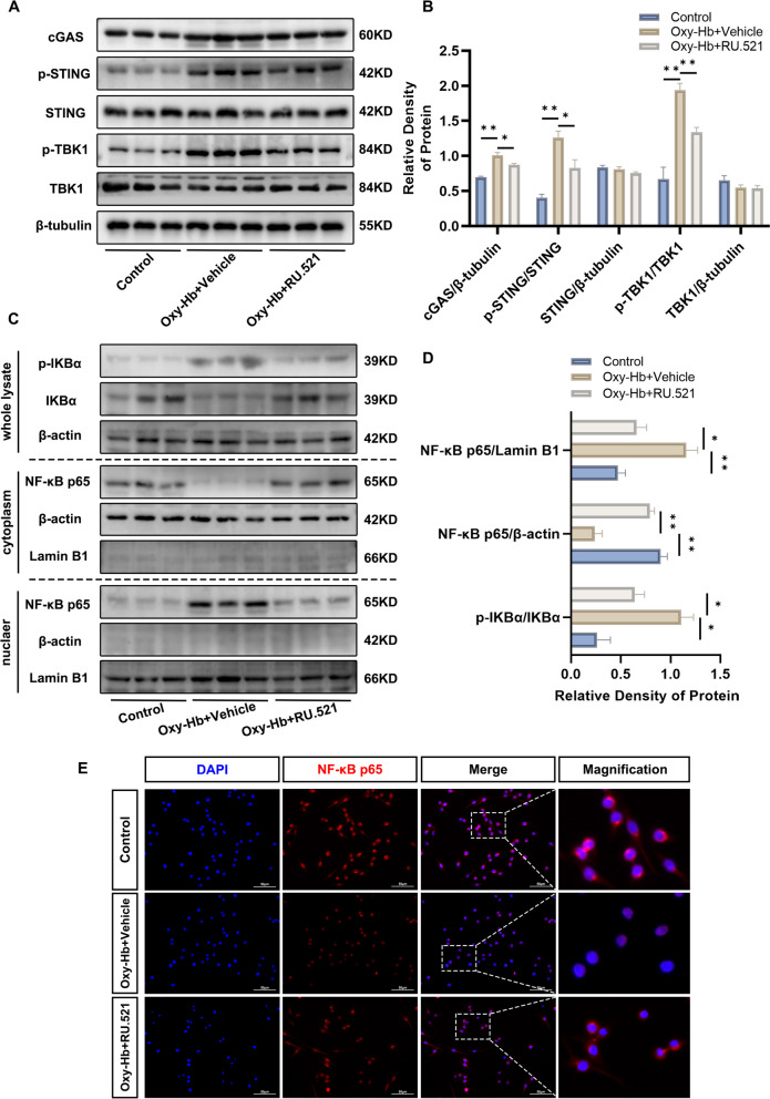
Methods: The expression and microglial localization of cGAS following SAH were investigated with western blot analysis and immunofluorescent double-staining, respectively. RU.521 was administered after SAH. 2'3'-cGAMP, a second messenger converted by activated cGAS, was used to activate cGAS-STING. The assessments were carried out by adopting various techniques including neurological function scores, brain water content, blood-brain barrier permeability, western blot analysis, TUNEL staining, Nissl staining, immunofluorescence, morphological analysis, Morris water maze test, Golgi staining, CCK8, flow cytometry in the in vivo and in vitro settings.

Results: Following SAH, there was an observed increase in the expression levels of cGAS in rat brain tissue, with peak levels observed at 24 h post-SAH. RU.521 resulted in a reduction of brain water content and blood-brain barrier permeability, leading to an improvement in neurological deficits after SAH. RU.521 had beneficial effects on neuronal apoptosis and microglia activation, as well as improvements in microglial morphology. Additionally, RU.521 prompted a shift in microglial phenotype from M1 to M2. We also noted a decrease in the production of pro-inflammatory cytokines TNF-α, IL-1β, and IL-6, and an increase in the level of the anti-inflammatory cytokine IL-10. Finally, RU.521 treatment was associated with improvements in cognitive function and an increase in the number of dendritic spines in the hippocampus. The therapeutic effects were mediated by the cGAS/STING/NF-κB pathway and were found to be abolished by 2'3'-cGAMP. In vitro, RU.521 significantly reduced apoptosis and neuroinflammation.

Conclusion: The study showed that SAH leads to neuroinflammation caused by microglial activation, which contributes to early brain injury. RU.521 improved neurological outcomes and reduced neuroinflammation by regulating microglial polarization through the cGAS/STING/NF-κB pathway in early brain injury after SAH. RU.521 may be a promising candidate for the treatment of neuroinflammatory pathology after SAH. Video Abstract.

Keywords: Early brain injury; Microglia; NF-κB; Neuroinflammation; STING; Subarachnoid hemorrhage; cGAS.


Conflict of interest statement

The authors declare no competing interests.

Figures

Fig. 1
Fig. 1
Expression changes of cGAS and microglial localization. A, B Representative Western blot bands of time course and quantitative analysis for cGAS. n = 6 per group. *: P < 0.05, **: P < 0.01, ***: P < 0.001, and ****: P < 0.0001. ns, not significant. C Representative images of double immunofluorescence staining for cGAS (green) with microglia (Iba1, red) at 24 h after SAH. Cell nuclei were counterstained with DAPI (blue). Scale bar = 50 μm. n = 6 per group
Fig. 2
Fig. 2
RU.521 was found to have a positive impact on neurological deficits, brain edema, BBB disruption, and neuronal damage 24 h after SAH. A, B The modified Garcia and beam balance scores of each group. n = 6 per group. C Quantification of brain water content at 24 h after SAH. n = 6 per group. D Quantification of Evans blue extravasation at 24 h after SAH. n = 6 per group. E Representative images of TUNEL staining (green) with neurons (NeuN, red). Scale bar = 100 μm. F Quantitative analysis of TUNEL-positive neuronal cells. n = 6 per group. G Representative images of Nissl staining. Scale bar = 100 μm. H Quantitative analysis of Nissl staining. n = 6 per group. *: P < 0.05, **: P < 0.01, ***: P < 0.001, and ****: P < 0.0001. ns, not significant
Fig. 3
Fig. 3
RU.521 blunted the over-activation of microglia after SAH. A, B Western blot bands and quantitative analysis for Iba1 expression. n = 6 per group. C, D Immunofluorescence staining and quantitative analysis of activated microglia (Iba1). Scale bar = 100 μm. n = 6 per group. E Representative images of activated microglia (green) for the measurement of morphological parameters and (F) quantitative analysis with the area, perimeter, process length, fractal dimension, lacunarity, density, span ratio, and circularity. Scale bar = 50 μm. n = 6 per group. *: P < 0.05, **: P < 0.01, ***: P < 0.001, and ****: P < 0.0001. ns, not significant
Fig. 4
Fig. 4
RU.521 prompted microglial M1-to-M2 polarization and attenuated neuroinflammation via the cGAS/STING/NF-κB pathway. A-D Western blot bands and quantitative analysis of the expression of p-STING, STING, p-TBK1, TBK1, p-NF-κB p65, NF-κB p65, Arg-1, CD16, IL-10, IL-6, TNF-α, and IL-1β. n = 6 per group. E The representative pictures of immunofluorescence and quantitative analysis for pro-inflammatory cytokine TNF-α. Scale bar = 50 μm. n = 6 per group. *: P < 0.05, **: P < 0.01, ***: P < 0.001, and ****: P < 0.0001. ns, not significant
Fig. 5
Fig. 5
RU.521 promoted the recovery of neurological function and dendritic spine densities. A, B Analysis of the modified Garcia scores and beam balance scores of each group. n = 6 per group. C, D Escape latency and swim distance in the visible platform trial of the water maze test. n = 6 per group. E Escape latency in the hidden platform trial of the water maze test. n = 6 per group. FH The times each animal crossed the original position of the platform, swimming speed, and swimming path in the probe trial of the water maze test. n = 6 per group. I, J Representative images of Golgi staining for dendritic spine densities and the analysis results. Scale bar = 2 μm. n = 6 per group. *: P < 0.05, **: P < 0.01, ***: P < 0.001, and ****: P < 0.0001. &: P < 0.05, &&: P < 0.01, &&&: P < 0.001, and &&&&: P < 0.0001. #: P < 0.05, ##: P < 0.01, ###: P < 0.001, and ####: P < 0.0001. ns, not significant
Fig. 6
Fig. 6
RU.521 inhibited neuronal damage in vitro. A Illustration of the co-culture system. B, D Representative western blot bands and quantitative analysis of the expression of cleaved-caspase 3 for TH22 cells. n = 3 per group. C, E Representative flow cytometry plots for HT22 cells. The apoptosis rate is calculated by the proportion of PI +/Annexin V+ plus PI /Annexin V+. n = 3 per group. F Quantitative analysis of cell viability detected by CCK8 assays. n = 3 per group. *: P < 0.05, **: P < 0.01, ***: P < 0.001, and ****: P < 0.0001. ns, not significant
Fig. 7
Fig. 7
RU.521 promoted microglial M1-to-M2 phenotypic polarization in vitro. A-C Western blot bands and quantitative analysis for target protein expression in BV2 cells. n = 3 per group. D-G Representative images of immunofluorescence staining and quantitative analysis for CD16, CD206, and TNF-α. Scale bar = 50/100 μm. n = 3 per group. *: P < 0.05, **: P < 0.01, ***: P < 0.001, and ****: P < 0.0001. ns, not significant
Fig. 8
Fig. 8
RU.521 inhibited microglial cGAS/STING pathway and the nuclear translocation of the p65 subunit of NF-κB in vitroA, B Immunoblots analysis for the expression of cGAS, p-STING, STING, p-TBK1, and TBK1. n = 3 per group. C, D Immunoblots and quantitative analysis of the expression of p-IKBα and NF-kB p65. n = 3 per group. E Immunofluorescence staining for NF-kB p65 (red) and cell nuclei with DAPI (blue). Scale bar = 50 μm. *: P < 0.05, **: P < 0.01, ***: P < 0.001, and ****: P < 0.0001. ns, not significant
Fig. 9
Fig. 9
Schematic illustration of the possible mechanisms. Briefly, the activation of the cGAS/STING/NF-κB pathway was induced by subarachnoid hemorrhage, resulting in the expression of inflammatory genes and microglial activation. This activation led microglia to polarize towards the M1 phenotype, resulting in neuroinflammation through increased production of pro-inflammatory cytokines, which could ultimately worsen brain injury. RU.521 can regulate microglial polarization and reduce neuroinflammation, thereby mitigating these harmful effects. The figure was created with Biorender.com. Agreement number: TW25N2UE2X

Similar articles

Cited by

References

    1. Lawton MT, Vates GE. Subarachnoid Hemorrhage. N Engl J Med. 2017;377:257–66. 10.1056/NEJMcp1605827 - DOI - PubMed
    1. Macdonald RL, Schweizer TA. Spontaneous subarachnoid haemorrhage. Lancet. 2017;389:655–66. 10.1016/S0140-6736(16)30668-7 - DOI - PubMed
    1. Zhang T, Wu P, Budbazar E, Zhu Q, Sun C, Mo J, Peng J, Gospodarev V, Tang J, Shi H, Zhang JH. Mitophagy Reduces Oxidative Stress Via Keap1 (Kelch-Like Epichlorohydrin-Associated Protein 1)/Nrf2 (Nuclear Factor-E2-Related Factor 2)/PHB2 (Prohibitin 2) Pathway After Subarachnoid Hemorrhage in Rats. Stroke. 2019;50:978–88. 10.1161/STROKEAHA.118.021590 - DOI - PMC - PubMed
    1. Li Y, Liu Y, Wu P, Tian Y, Liu B, Wang J, Bihl J, Shi H. Inhibition of Ferroptosis alleviates early brain injury after subarachnoid hemorrhage in vitro and in vivo via reduction of lipid peroxidation. Cell Mol Neurobiol. 2021;41:263–78. 10.1007/s10571-020-00850-1 - DOI - PMC - PubMed
    1. Tian Y, Liu B, Li Y, Zhang Y, Shao J, Wu P, Xu C, Chen G, Shi H. Activation of RARalpha Receptor Attenuates Neuroinflammation After SAH via Promoting M1-to-M2 Phenotypic Polarization of Microglia and Regulating Mafb/Msr1/PI3K-Akt/NF-kappaB Pathway. Front Immunol. 2022;13:839796. 10.3389/fimmu.2022.839796 - DOI - PMC - PubMed

Publication types