

Challenges of aortic valve tissue culture - maintenance of viability and extracellular matrix in the pulsatile dynamic microphysiological system
- PMID: 37770970
- PMCID: PMC10538250
- DOI: 10.1186/s13036-023-00377-1
Challenges of aortic valve tissue culture - maintenance of viability and extracellular matrix in the pulsatile dynamic microphysiological system
Abstract
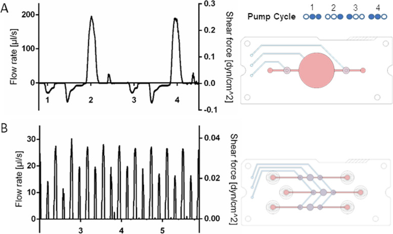
Background: Calcific aortic valve disease (CAVD) causes an increasing health burden in the 21st century due to aging population. The complex pathophysiology remains to be understood to develop novel prevention and treatment strategies. Microphysiological systems (MPSs), also known as organ-on-chip or lab-on-a-chip systems, proved promising in bridging in vitro and in vivo approaches by applying integer AV tissue and modelling biomechanical microenvironment. This study introduces a novel MPS comprising different micropumps in conjunction with a tissue-incubation-chamber (TIC) for long-term porcine and human AV incubation (pAV, hAV).
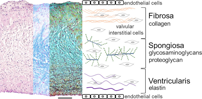
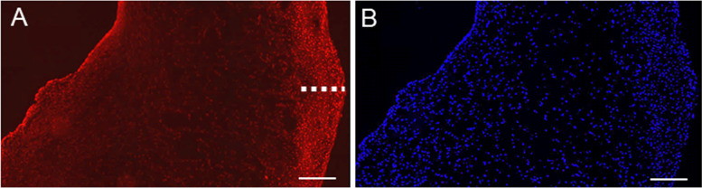
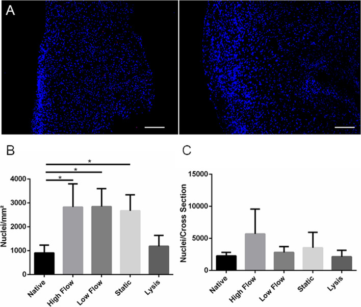
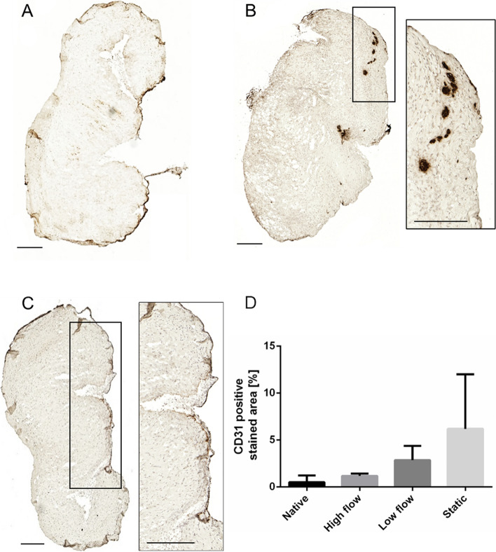
Results: Tissue cultures in two different MPS setups were compared and validated by a bimodal viability analysis and extracellular matrix transformation assessment. The MPS-TIC conjunction proved applicable for incubation periods of 14-26 days. An increased metabolic rate was detected for pulsatile dynamic MPS culture compared to static condition indicated by increased LDH intensity. ECM changes such as an increase of collagen fibre content in line with tissue contraction and mass reduction, also observed in early CAVD, were detected in MPS-TIC culture, as well as an increase of collagen fibre content. Glycosaminoglycans remained stable, no significant alterations of α-SMA or CD31 epitopes and no accumulation of calciumhydroxyapatite were observed after 14 days of incubation.
Conclusions: The presented ex vivo MPS allows long-term AV tissue incubation and will be adopted for future investigation of CAVD pathophysiology, also implementing human tissues. The bimodal viability assessment and ECM analyses approve reliability of ex vivo CAVD investigation and comparability of parallel tissue segments with different treatment strategies regarding the AV (patho)physiology.
Keywords: Calcific aortic valve disease; ECM remodelling; Microphysiological system; Tissue culture; Viability.
© 2023. BioMed Central Ltd., part of Springer Nature.
Conflict of interest statement
The authors declare no competing interests.
Figures


Similar articles
-
Establishment of a resazurin-based aortic valve tissue viability assay for dynamic culture in a microphysiological system.Clin Hemorheol Microcirc. 2021;79(1):167-178. doi: 10.3233/CH-219112. Clin Hemorheol Microcirc. 2021. PMID: 34487029
-
Reproducible In Vitro Tissue Culture Model to Study Basic Mechanisms of Calcific Aortic Valve Disease: Comparative Analysis to Valvular Interstitials Cells.Biomedicines. 2021 Apr 26;9(5):474. doi: 10.3390/biomedicines9050474. Biomedicines. 2021. PMID: 33925890 Free PMC article.
-
Characterizing the reproducibility in using a liver microphysiological system for assaying drug toxicity, metabolism, and accumulation.Clin Transl Sci. 2021 May;14(3):1049-1061. doi: 10.1111/cts.12969. Epub 2021 Apr 3. Clin Transl Sci. 2021. PMID: 33382907 Free PMC article.
-
Insights into calcific aortic valve stenosis: a comprehensive overview of the disease and advancing treatment strategies.Ann Med Surg (Lond). 2024 Apr 29;86(6):3577-3590. doi: 10.1097/MS9.0000000000002106. eCollection 2024 Jun. Ann Med Surg (Lond). 2024. PMID: 38846838 Free PMC article. Review.
-
Impact of calcific aortic valve disease on valve mechanics.Biomech Model Mechanobiol. 2022 Feb;21(1):55-77. doi: 10.1007/s10237-021-01527-4. Epub 2021 Oct 23. Biomech Model Mechanobiol. 2022. PMID: 34687365 Review.
Cited by
-
Oxygenator assisted dynamic microphysiological culture elucidates the impact of hypoxia on valvular interstitial cell calcification.J Biol Eng. 2024 Aug 23;18(1):45. doi: 10.1186/s13036-024-00441-4. J Biol Eng. 2024. PMID: 39180097 Free PMC article.
References
-
- Arastéh K, Baenkler H, Bieber C. Innere Medizin [Internet]. Reihe D, editor. Innere Medizinpie. Stuttgart: Georg Thieme Verlag; 2018. p. 131–155. Available from: https://eref.thieme.de/10.1055/b-005-145255. - DOI
-
- Coffey S, Roberts-Thomson R, Brown A, Carapetis J, Chen M, Enriquez-Sarano M, et al. Global epidemiology of valvular heart disease. Nat Rev Cardiol. 2021;18(12):853–864. - PubMed
-
- Fernandez Esmerats J, Villa-Roel N, Kumar S, Gu L, Salim MT, Ohh M, et al. Disturbed flow increases UBE2C (Ubiquitin E2 Ligase C) via Loss of miR-483-3p, inducing aortic valve calcification by the pVHL (von Hippel-Lindau Protein) and HIF-1α (Hypoxia-Inducible Factor-1α) pathway in endothelial cells. Arterioscler Thromb Vasc Biol. 2019;39(3):467–481. - PMC - PubMed
Grants and funding
LinkOut - more resources
Full Text Sources
Miscellaneous
