

Synchrotron microtomography reveals insights into the degradation kinetics of bio-degradable coronary magnesium scaffolds
- PMID: 37771679
- PMCID: PMC10522944
- DOI: 10.1016/j.bioactmat.2023.09.008
Synchrotron microtomography reveals insights into the degradation kinetics of bio-degradable coronary magnesium scaffolds
Abstract
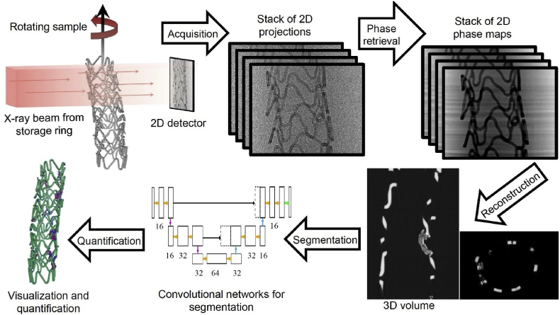
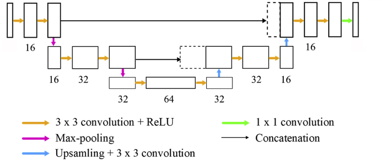
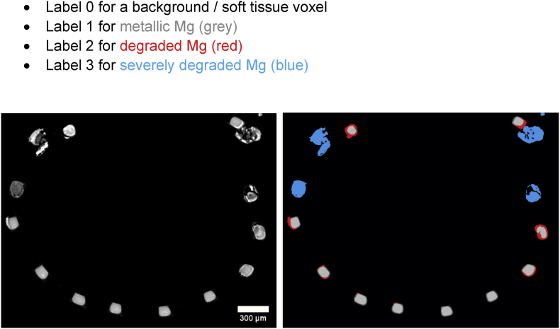
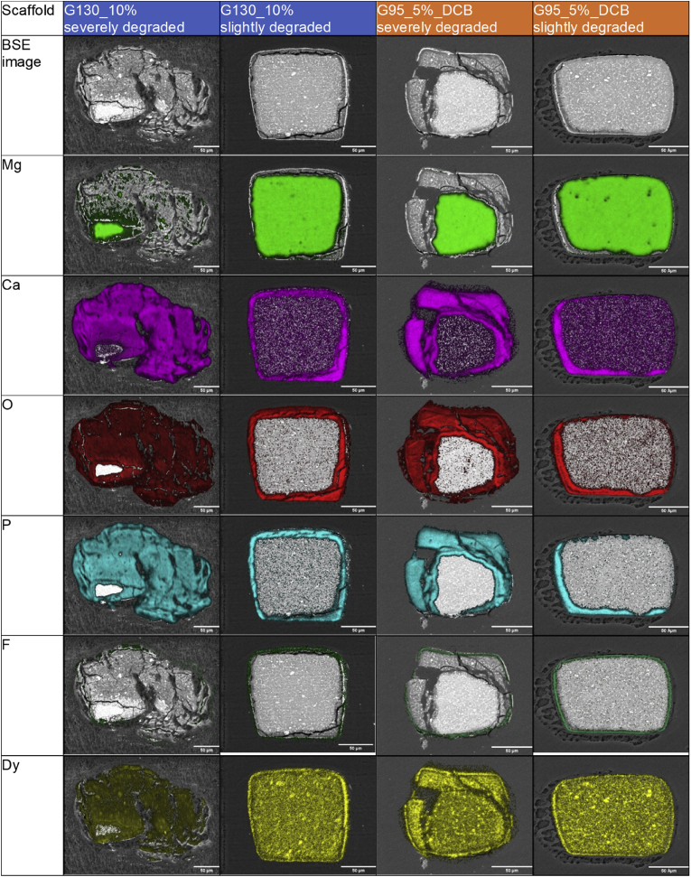
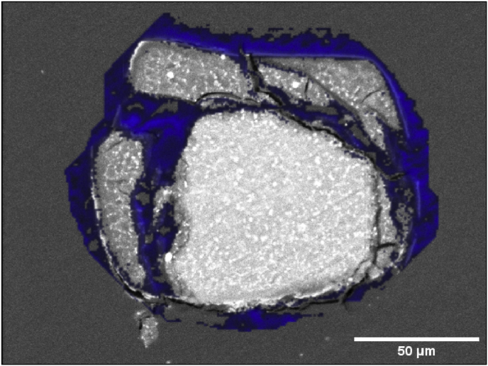
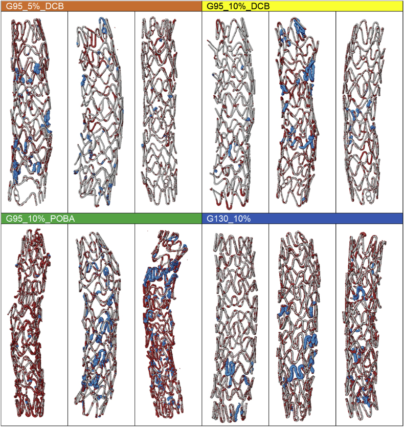
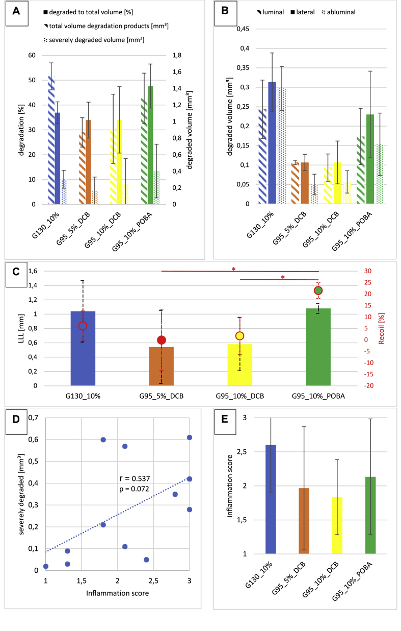
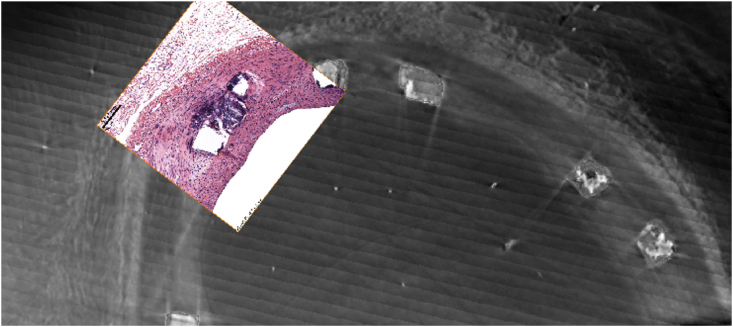
Bioresorbable magnesium scaffolds are a promising future treatment option for coronary artery stenosis, especially for young adults. Due to the degradation of these scaffolds (<1 year), long-term device-related clinical events could be reduced compared to treatments with conventional drug eluting stents. First clinical trials indicate a return of vasomotion after one year, which may be associated with improved long-term clinical outcomes. However, even after decades of development, the degradation process, ideal degradation time and biological response in vivo are still not fully understood. The present study investigates the in vivo degradation of magnesium scaffolds in the coronary arteries of pigs influenced by different strut thicknesses and the presence of antiproliferative drugs. Due to high 3D image contrast of synchrotron-based micro-CT with phase contrast (SR-μCT), a qualitative and quantitative evaluation of the degradation morphology of magnesium scaffolds was obtained. For the segmentation of the μCT images a convolutional network architecture (U-net) was exploited, demonstrating the huge potential of merging high resolution SR-μCT with deep learning (DL) supported data analysis. In total, 30 scaffolds, made of the rare earth alloy Resoloy®, with different strut designs were implanted into the coronary arteries of 10 domestic pigs for 28 days using drug-coated or uncoated angioplasty balloons for post-dilatation. The degradation morphology was analyzed using scanning electron microscopy, energy dispersive x-ray spectroscopy and SR-μCT. The data from these methods were then related to data from angiography, optical coherence tomography and histology. A thinner strut size (95 vs. 130 μm) and the presence of paclitaxel indicated a slower degradation rate at 28 d in vivo, which positively influences the late lumen loss (0.5 and 0.6 mm vs. 1.0 and 1.1 mm) and recoil values (0 and 1.7% vs. 6.1 and 22%).
Keywords: Artificial intelligence; DCB; Degradation; Inflammation; Magnesium scaffold; Paclitaxel; Resoloy.
© 2023 The Authors.
Conflict of interest statement
R. Menze is an employee of MeKo Manufacturing e.K., B. Hesse is CEO and shareholder of Xploraytion GmbH, D. Chen was an employee of Xploraytion GmbH, M. Kusmierczuk is an employee and B. Scheller is a shareholder of InnoRa GmbH, T. Weitkamp and S. Bettink have no conflicts of interest to declare.
Figures
References
-
- Lipinski M.J., Acampado E., Cheng Q., Adams L., Torii S., Gai J., Torguson R., Hellinga D.G., Joner M., Harder C., Zumstein P., Finn A.V., Kolodgie F.D., Virmani R., Waksman R. Comparison of acute thrombogenicity for magnesium versus stainless steel stents in a porcine arteriovenous shunt model. EuroIntervention. 2019;14 1420–7. - PubMed
-
- Joner M., Ruppelt P., Zumstein P., Lapointe-Corriveau C., Leclerc G., Bulin A., Castellanos M.I., Wittchow E., Haude M., Waksman R. Preclinical evaluation of degradation kinetics and elemental mapping of first- and second-generation bioresorbable magnesium scaffolds. EuroIntervention. 2018;14:e1040–e1048. - PubMed
-
- Serruys P.W., Chevalier B., Sotomi Y., Cequier A., Carrié D., Piek J.J., Van Boven A.J., Dominici M., Dudek D., McClean D., Helqvist S., Haude M., Reith S., de Sousa Almeida M., Campo G., Iñiguez A., Sabaté M., Windecker S., Onuma Y. Comparison of an everolimus-eluting bioresorbable scaffold with an everolimus-eluting metallic stent for the treatment of coronary artery stenosis (ABSORB II): a 3 year, randomised, controlled, single-blind, multicentre clinical trial. Lancet. 2016;388:2479–2491. - PubMed
-
- Verheye S., Wlodarczak A., Montorsi P., Torzewski J., Bennett J., Haude M., Starmer G., Buck T., Wiemer M., Nuruddin A.A.B., Yan B.P.Y., Lee M.K.Y. Catheter Cardiovasc Interv; 2020. BIOSOLVE-IV-registry: Safety and Performance of the Magmaris Scaffold: 12-month Outcomes of the First Cohort of 1,075 Patients. - PMC - PubMed
-
- Haude M., Toelg R., Lemos P.A., Christiansen E.H., Abizaid A., von Birgelen C., Neumann F.-J., Wijns W., Ince H., Kaiser C., Lim S.T., Escaned J., Eeckhout E., Garcia-Garcia H.M., Waksman R. Sustained safety and performance of a second-generation sirolimus-eluting absorbable metal scaffold: long-term data of the BIOSOLVE-II first-in-man trial at 5 years. Cardiovasc. Revascularization Med. 2022;38:106–110. - PubMed
LinkOut - more resources
Full Text Sources
Research Materials
