

Evaluation of CI electrode position from imaging: comparison of an automated technique with the established manual method
- PMID: 37773060
- PMCID: PMC10543862
- DOI: 10.1186/s12880-023-01102-6
Evaluation of CI electrode position from imaging: comparison of an automated technique with the established manual method
Abstract
Background: A manual evaluation of the CI electrode position from CT and DVT scans may be affected by diagnostic errors due to cognitive biases. The aim of this study was to compare the CI electrode localization using an automated method (image-guided cochlear implant programming, IGCIP) with the clinically established manual method.
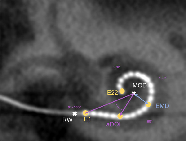
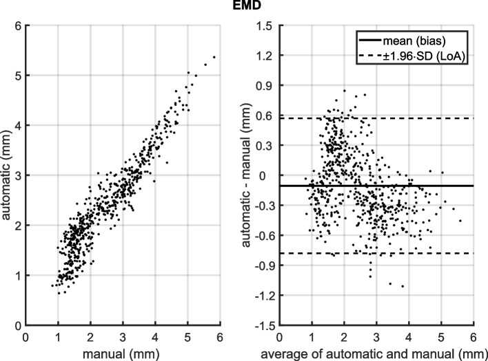
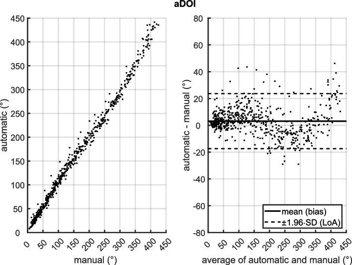
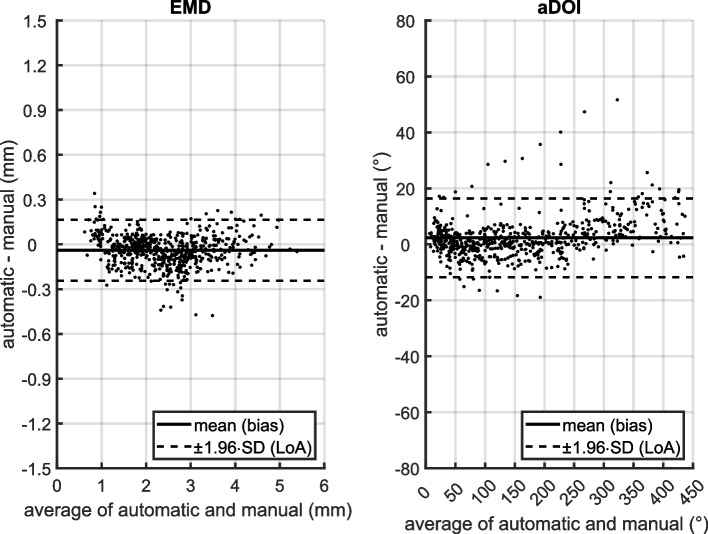
Methods: This prospective experimental study was conducted on a dataset comprising N=50 subjects undergoing cochlear implantation with a Nucleus® CI532 or CI632 Slim Modiolar electrode. Scalar localization, electrode-to-modiolar axis distances (EMD) and angular insertion depth (aDOI) were compared between the automated IGCIP tool and the manual method. Two raters made the manual measurements, and the interrater reliability (±1.96·SD) was determined as the reference for the method comparison. The method comparison was performed using a correlation analysis and a Bland-Altman analysis.
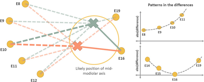
Results: Concerning the scalar localization, all electrodes were localized both manually and automatically in the scala tympani. The interrater differences ranged between ±0.2 mm (EMD) and ±10° (aDOI). There was a bias between the automatic and manual method in measuring both localization parameters, which on the one hand was smaller than the interrater variations. On the other hand, this bias depended on the magnitude of the EMD respectively aDOI. A post-hoc analysis revealed that the deviations between the methods were likely due to a different selection of mid-modiolar axis.
Conclusions: The IGCIP is a promising tool for automated processing of CT and DVT scans and has useful functionality such as being able to segment the cochlear using post-operative scans. When measuring EMD, the IGCIP tool is superior to the manual method because the smallest possible distance to the axis is determined depending on the cochlear turn, whereas the manual method selects the helicotrema as the reference point rigidly. Functionality to deal with motion artifacts and measurements of aDOI according to the consensus approach are necessary, otherwise the IGCIP is not unrestrictedly ready for clinical use.
Keywords: Angular depth of insertion; Cochlear implant; EMD; Electrode localization; Electrode-to-modiolus-distance; IGCIP; aDOI; imaging.
© 2023. BioMed Central Ltd., part of Springer Nature.
Conflict of interest statement
AM, JD, GB and MH declare general financial support by Cochlear Ltd. (research grant to institution). CB is an employee of Cochlear Ltd. (Sydney, Australia). CB contributed to the data collection, analysis, and preparation of this manuscript. Cochlear Ltd. was not involved in the study design and decision to publish. The authors alone are responsible for the content and writing of this paper. JD receives financial support within the framework of the clinician-scientist program of Kiel University’s faculty of medicine.
Figures

Similar articles
-
Quality-assured training in the evaluation of cochlear implant electrode position: a prospective experimental study.BMC Med Educ. 2022 May 20;22(1):386. doi: 10.1186/s12909-022-03464-x. BMC Med Educ. 2022. PMID: 35596162 Free PMC article.
-
Clinical investigation of the Nucleus Slim Modiolar Electrode.Audiol Neurootol. 2017;22(3):169-179. doi: 10.1159/000480345. Epub 2017 Oct 24. Audiol Neurootol. 2017. PMID: 29059669
-
Comparison of Skull Radiograph and Computed Tomography Measurements of Cochlear Implant Insertion Angles.Otol Neurotol. 2019 Mar;40(3):e298-e303. doi: 10.1097/MAO.0000000000002121. Otol Neurotol. 2019. PMID: 30741910 Free PMC article.
-
Automated Calculation of Cochlear Implant Electrode Insertion Parameters in Clinical Cone-Beam CT.Otol Neurotol. 2022 Feb 1;43(2):199-205. doi: 10.1097/MAO.0000000000003432. Otol Neurotol. 2022. PMID: 34789695
-
Cochlear Implant Electrode Tip Fold-Over: Our Experience With Long and Flexible Electrode.Otol Neurotol. 2022 Jan 1;43(1):64-71. doi: 10.1097/MAO.0000000000003362. Otol Neurotol. 2022. PMID: 34619728 Review.
Cited by
-
Curvature analysis of CI electrode arrays: a novel approach to categorize perimodiolar positions without anatomical landmarks.Eur Arch Otorhinolaryngol. 2025 Jan;282(1):145-154. doi: 10.1007/s00405-024-08917-1. Epub 2024 Aug 30. Eur Arch Otorhinolaryngol. 2025. PMID: 39214908 Free PMC article.
References
Publication types
MeSH terms
LinkOut - more resources
Full Text Sources
Medical
Research Materials
