. 2023 Sep 29;27(1):374.
doi: 10.1186/s13054-023-04652-x.

Therapeutic S100A8/A9 blockade inhibits myocardial and systemic inflammation and mitigates sepsis-induced myocardial dysfunction

Affiliations

Therapeutic S100A8/A9 blockade inhibits myocardial and systemic inflammation and mitigates sepsis-induced myocardial dysfunction

Gabriel Jakobsson et al. Crit Care. .

Abstract

Background and aims: The triggering factors of sepsis-induced myocardial dysfunction (SIMD) are poorly understood and are not addressed by current treatments. S100A8/A9 is a pro-inflammatory alarmin abundantly secreted by activated neutrophils during infection and inflammation. We investigated the efficacy of S100A8/A9 blockade as a potential new treatment in SIMD.

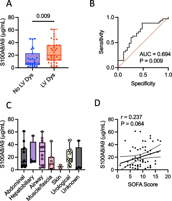
Methods: The relationship between plasma S100A8/A9 and cardiac dysfunction was assessed in a cohort of 62 patients with severe sepsis admitted to the intensive care unit of Linköping University Hospital, Sweden. We used S100A8/A9 blockade with the small-molecule inhibitor ABR-238901 and S100A9-/- mice for therapeutic and mechanistic studies on endotoxemia-induced cardiac dysfunction in mice.

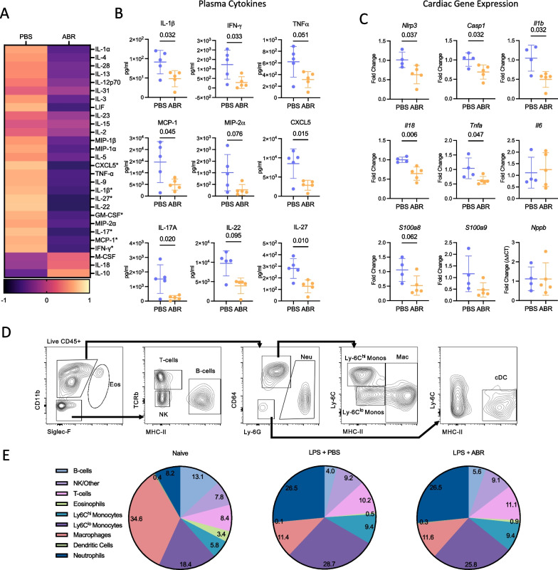
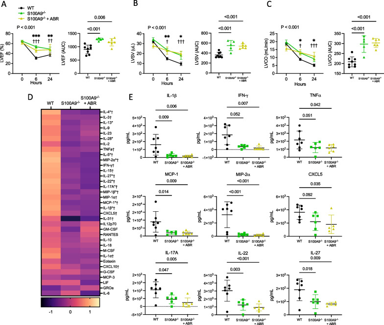
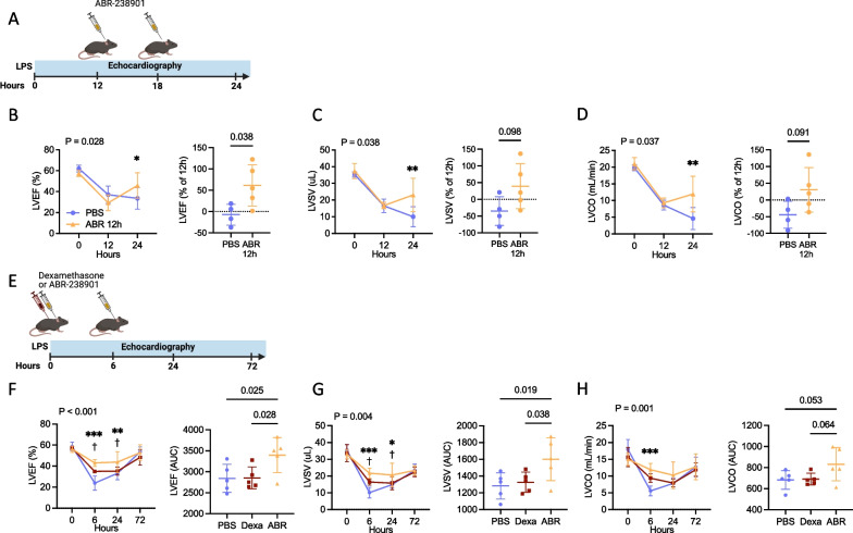
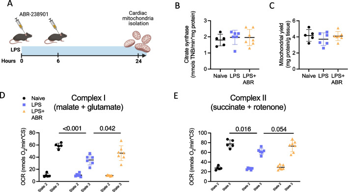
Results: In sepsis patients, elevated plasma S100A8/A9 was associated with left-ventricular (LV) systolic dysfunction and increased SOFA score. In wild-type mice, 5 mg/kg of bacterial lipopolysaccharide (LPS) induced rapid plasma S100A8/A9 increase and acute LV dysfunction. Two ABR-238901 doses (30 mg/kg) administered intraperitoneally with a 6 h interval, starting directly after LPS or at a later time-point when LV dysfunction is fully established, efficiently prevented and reversed the phenotype, respectively. In contrast, dexamethasone did not improve cardiac function compared to PBS-treated endotoxemic controls. S100A8/A9 inhibition potently reduced systemic levels of inflammatory mediators, prevented upregulation of inflammatory genes and restored mitochondrial function in the myocardium. The S100A9-/- mice were protected against LPS-induced LV dysfunction to an extent comparable with pharmacologic S100A8/A9 blockade. The ABR-238901 treatment did not induce an additional improvement of LV function in the S100A9-/- mice, confirming target specificity.

Conclusion: Elevated S100A8/A9 is associated with the development of LV dysfunction in severe sepsis patients and in a mouse model of endotoxemia. Pharmacological blockade of S100A8/A9 with ABR-238901 has potent anti-inflammatory effects, mitigates myocardial dysfunction and might represent a novel therapeutic strategy for patients with severe sepsis.

Keywords: Endotoxemia; Inflammation; Mitochondrial function; Neutrophils; S100A8/A9; Sepsis-induced myocardial dysfunction.


Conflict of interest statement

Michelle Chew is a member of the editorial board of Critical Care.

Figures

Fig. 1
Fig. 1
S100A8/A9 is elevated in critically ill sepsis patients with left ventricular dysfunction. S100A8/A9 was measured in plasma collected upon admittance into the ICU. A Comparison of plasma S100A8/A9 levels in patients with severe sepsis, with or without left ventricular dysfunction (LV Dys). B ROC analysis of the ability of S100A8/A9 at admission to identify LV Dys. C Relationship between S100A8/A9 levels and the site of infection. D Correlation between plasma S100A8/A9 and SOFA disease severity score. Comparison of S100A8/A9 in patients with or without LV Dys was performed using the Mann–Whitney U test. Spearman correlation analysis was used to assess the relationship between S100A8/A9 and the SOFA score
Fig. 2
Fig. 2
S100A8/A9 blockade ameliorates septic cardiomyopathy. Endotoxemia was induced in C57Bl/6NrJ mice by intraperitoneal injection of 5 mg/kg LPS. The mice were treated with 30 mg/kg ABR-238901 (ABR) or PBS intraperitoneally at 0 h and 6 h post-LPS. A Experimental timeline for BC. Blood sampling was performed at 0, 6, 12, 24, 48, 72 and 96 h post-LPS. B Kinetics of S100A8/A9 release. C Bodyweight change, expressed as serial measurements and area under the curve (AUC). D Experimental timeline for EI. Echocardiography was performed at baseline, 6 h, 12 h and 24 h. EG Left ventricular ejection fraction (LVEF), stroke volume (LVSV) and cardiac output (LVCO), presented as serial measurements and area under the curve over time. Statistical testing was performed by repeated-measures two-way ANOVA with Fisher’s LSD Test. The p-values on the time-course graphs reflect differences between the treatment groups at the respective time point. Differences in AUC between the groups were assessed using Student’s t-test, following normality assessment with Shapiro–Wilk test. PBS, Phosphate Buffered Saline; ABR, ABR-238901; BW, bodyweight; AUC, area under curve; LVEF, Left ventricular ejection fraction; LVSV, Left ventricular stroke volume; LVCO, Left ventricular cardiac output. Data is represented as mean ± SD. B and C N = 7–15 per group, EG N = 5 per group
Fig. 3
Fig. 3
S100A8/A9 blockade inhibits systemic inflammation but does not impact cardiac immune cell environment during endotoxemia. Endotoxemia was induced in C57Bl/6NrJ mice by intraperitoneal injection of 5 mg/kg LPS. The mice were treated with 30 mg/kg ABR-238901 or PBS intraperitoneally at 0 h and 6 h post-LPS. The mice were sacrificed at 24 h for plasma isolation and flow cytometric analysis (A, B, D, E), or at 12 h for cardiac gene expression (C). Cytokine levels were measured in plasma. A Heatmap of plasma cytokine changes following ABR-238901 treatment during endotoxemia, expressed as Z-score in relation to median. B Treatment effect on plasma levels of individual pro-inflammatory cytokines and chemokines. C Cardiac gene expression of NLRP3 inflammasome components, inflammatory cytokines, S100A8, S100A9 and BNP. D Gating strategy for flow cytometry analysis of cardiac CD45+ leukocytes. E Distribution of cardiac immune cell populations during endotoxemia, with and without S100A8/A9 blockade. Statistical differences between two groups were examined with Student’s t-test or Mann–Whitney test, following normality assessment with Shapiro–Wilk test. *P < 0.05, PBS, Phosphate Buffered Saline; ABR, ABR-238901; Eos, Eosinophils; NK, Natural Killer cells; Neu, Neutrophils; Monos, Monocytes; Mac, Macrophages; cDC, conventional dendritic cells. Data is represented as mean ± SD. A and B N = 5 per group, C N = 4-5 per group, D and E N = 5–8 per group
Fig. 4
Fig. 4
S100A9-deficient mice are partially protected against systemic inflammation and left ventricular dysfunction during endotoxemia. Endotoxemia was induced in wild-type or S100A9−/− C57Bl/6NrJ mice by intraperitoneal injection of 5 mg/kg LPS. In the ABR-238901-treated group, the S100A9−/− mice received 30 mg/kg ABR-238901 intraperitoneally at 0h and 6h post-LPS. Echocardiography was performed at baseline, 6 h and 24 h. Mice were sacrificed at 24 h for plasma collection and cytokines were measured in plasma using multiplex bead assay. AC Left ventricular ejection fraction, stroke volume and cardiac output presented as serial measurements over time, with the corresponding area under the curve (AUC). D Heatmap of changes in plasma cytokines in wild-type and S100A9−/− mice with and without ABR-238901 treatment, expressed as Z-scores. E Individual plasma cytokine and chemokine levels. Statistical testing of echocardiographic parameters over time was performed using repeated-measures 2-way ANOVA with Fisher’s LSD Test. P-values reflect differences between treatment groups over time. Symbols reflect the difference between the treatment groups at the respective time point. Statistical differences between 3 groups were assessed with 1-way ANOVA with Fisher’s LSD Test or Kruskal–Wallis test, following normality assessment with Shapiro–Wilk test. *, S100A9−/− versus WT; *P < 0.05, **P < 0.01, ***P < 0.001; †, S100A9−/− + ABR versus WT; † P < 0.05, †† P < 0.01, ††† P < 0.001; WT, wild-type; ABR, ABR-238901; AUC, area under curve; LVEF, Left ventricular ejection fraction; LVSV, Left ventricular stroke volume; LVCO, Left ventricular cardiac output. Data is represented as mean ± SD. N = 5–8 per group
Fig. 5
Fig. 5
ABR-238901 reverses established cardiac dysfunction and is a more efficient treatment compared to Dexamethasone. AE Delayed ABR-238901 treatment starting from 12 h post LPS successfully rescues cardiac dysfunction during endotoxemia. A Experimental layout for BD. B Repeated LVEF measurements and % LVEF change from treatment start to 24h post-LPS. C Absolute LVSV and % LVSV change from treatment start. D Absolute LVCO and % LVCO change from treatment start. E Experimental layout for FH. FH Comparison between treatment with ABR-238901 and Dexamethasone on cardiac function during endotoxemia. Following disease induction, the mice were treated either with 2 mg/kg Dexamethasone at 0h or with ABR-238901 at 0 and 6 h post-LPS. FH LVEF, LVSV and LVCO over time and area under the curve. Statistical testing of echocardiographic parameters over time was performed by repeated-measures 2-way ANOVA with Fisher’s LSD Test. The P-values reflect the difference between treatment groups over time. The symbols reflect the difference between treatment groups at the respective time point. The statistical difference between two groups was tested with Student’s t-test. Differences between three groups were tested using 1-way ANOVA with Fisher’s LSD Test. Normality assessment was performed with Shapiro–Wilk test. *, ABR versus PBS; *P < 0.05, **P < 0.01, ***P < 0.001; †, ABR vs Dexa; † P < 0.05; PBS, Phosphate Buffered Saline; ABR, ABR-238901; Dexa, Dexamethasone; AUC, area under curve; LVEF, Left ventricular ejection fraction; LVSV, Left ventricular stroke volume; LVCO, Left ventricular cardiac output. Data is presented as mean ± SD. N = 4–5 per group
Fig. 6
Fig. 6
S100A8/A9 blockade improves LPS-induced mitochondrial dysfunction in the heart. A Experimental layout. B Citrate synthase activity. C Mitochondrial yield. D Oxygen consumption rate (OCR) after incubation with respiratory substrates (malate + glutamate) feeding complex I, either in the absence (state 2) or presence (state 3) of ADP. E Oxygen consumption rate after incubation with respiratory substrates (succinate + rotenone) feeding complex II. Differences between three groups were tested using 1-way ANOVA with Fisher’s LSD Test. Normality assessment was performed with Shapiro–Wilk test. LPS, Lipopolysaccharide; ABR, ABR-238901; OCR, Oxygen consumption rate. Data are represented as mean ± SD from N = 4–7 per group

Comment in

References

    1. Gotts JE, Matthay MA. Sepsis: pathophysiology and clinical management. BMJ. 2016;353:i1585. - PubMed
    1. Angus DC, van der Poll T. Severe sepsis and septic shock. New Engl J Med. 2013;369:840–851. - PubMed
    1. Beesley SJ, Weber G, Sarge T, Nikravan S, Grissom CK, Lanspa MJ, et al. Septic cardiomyopathy. Crit Care Med. 2018;46:625–634. - PubMed
    1. Calvin JE, Driedger AA, Sibbald WJ. An assessment of myocardial function in human sepsis utilizing ECG gated cardiac scintigraphy. Chest. 1981;80:579–586. - PubMed
    1. L’Heureux M, Sternberg M, Brath L, Turlington J, Kashiouris MG. Sepsis-induced cardiomyopathy: a comprehensive review. Curr Cardiol Rep. 2020;22:35. - PMC - PubMed