

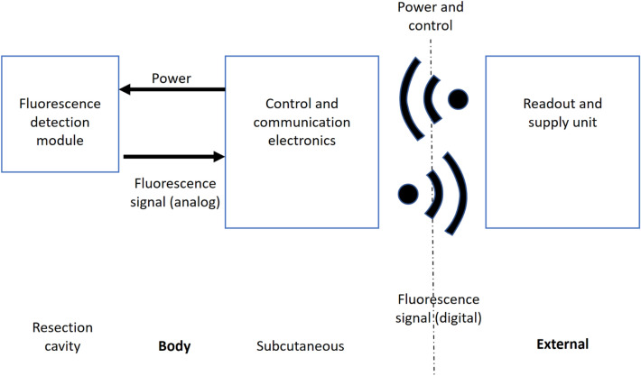
Concept of a fully-implantable system to monitor tumor recurrence
- PMID: 37773315
- PMCID: PMC10541913
- DOI: 10.1038/s41598-023-43226-3
Concept of a fully-implantable system to monitor tumor recurrence
Abstract
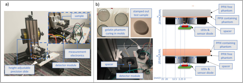
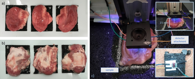
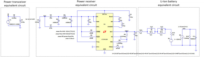
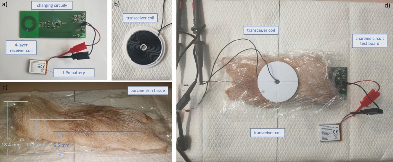
Current treatment for glioblastoma includes tumor resection followed by radiation, chemotherapy, and periodic post-operative examinations. Despite combination therapies, patients face a poor prognosis and eventual recurrence, which often occurs at the resection site. With standard MRI imaging surveillance, histologic changes may be overlooked or misinterpreted, leading to erroneous conclusions about the course of adjuvant therapy and subsequent interventions. To address these challenges, we propose an implantable system for accurate continuous recurrence monitoring that employs optical sensing of fluorescently labeled cancer cells and is implanted in the resection cavity during the final stage of tumor resection. We demonstrate the feasibility of the sensing principle using miniaturized system components, optical tissue phantoms, and porcine brain tissue in a series of experimental trials. Subsequently, the system electronics are extended to include circuitry for wireless energy transfer and power management and verified through electromagnetic field, circuit simulations and test of an evaluation board. Finally, a holistic conceptual system design is presented and visualized. This novel approach to monitor glioblastoma patients is intended to early detect recurrent cancerous tissue and enable personalization and optimization of therapy thus potentially improving overall prognosis.
© 2023. Springer Nature Limited.
Conflict of interest statement
The authors declare no competing interests.
Figures


References
-
- Figge, F. & Weiland, G. The affinity of neoplastic, embryonic, and traumatized tissue for porphyrins and metalloporphyrins. In Anatomical record100, 659–659, (Wiley-liss div john wiley & Sons Inc, 1948). - PubMed
Publication types
MeSH terms
LinkOut - more resources
Full Text Sources
