. 2023 Sep 29;13(1):16379.
doi: 10.1038/s41598-023-43518-8.

Choroidal morphology and microvascular structure in eyes of patients with idiopathic normal pressure hydrocephalus before and after ventriculo-peritoneal shunt surgery

Affiliations

Choroidal morphology and microvascular structure in eyes of patients with idiopathic normal pressure hydrocephalus before and after ventriculo-peritoneal shunt surgery

Nicola Valsecchi et al. Sci Rep. .

Abstract

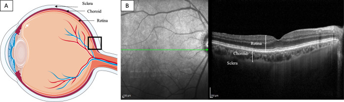
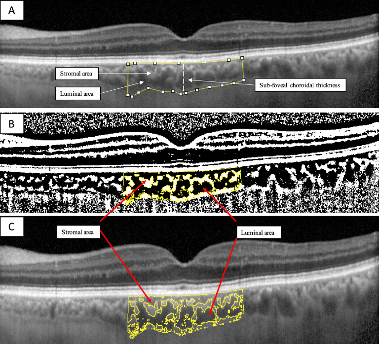
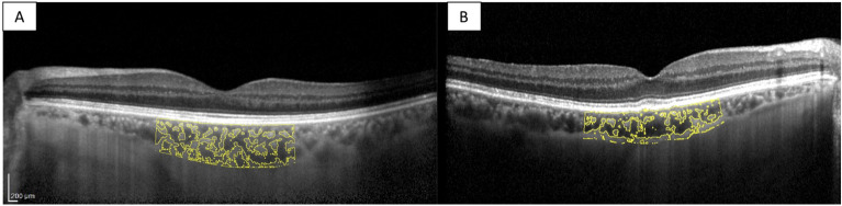
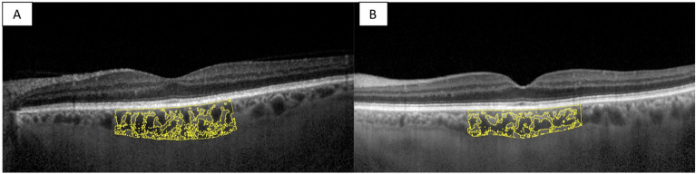
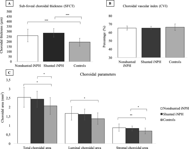
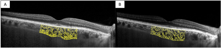
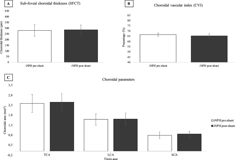
The present study aims to investigate the choroidal morphology and microvascular structure in eyes of patients with idiopathic normal pressure hydrocephalus (iNPH) compared with the eyes of healthy age-matched individuals, and to assess the choroidal structure in eyes of iNPH patients before and after shunt surgery using Optical Coherence Tomography (OCT). The primary objective was to assess the choroidal morphology in eyes of iNPH patients before and after ventriculo-peritoneal (VP) surgery compared to age and sex-matched healthy individuals. The secondary objective was to compare the choroidal morphology of iNPH patients before and after a mean of 56 days from shunt surgery. Eighteen consecutive patients diagnosed with iNPH and 18 healthy controls were prospectively recruited between November 2021 and October 2022. Spectral-domain optical coherence tomography (SD-OCT) with enhanced depth imaging (EDI) was conducted before and within 4 months after shunt surgery. Images were binarized using the ImageJ software, and the choroidal vascular index (CVI) was calculated. Sub-foveal choroidal thickness (SFCT), total choroidal area (TCA), luminal choroidal area (LCA), and stromal choroidal area (SCA) were significantly increased in iNPH patients before surgery compared to the control group (p < 0.05). SFCT, TCA, and SCA were significantly increased in iNPH patients after surgery compared to the control group (p < 0.05). There were no differences in the CVI between iNPH patients and controls. No statistical differences in the choroidal structure were observed before and after VP shunt surgery (p > 0.05). In conclusion, the choroid was thicker in iNPH patients before and after VP shunt compared to age-matched healthy individuals. However, there were no difference in the choroidal microstructure in the eyes of iNPH patients before and after a mean of 3 months from VP shunt surgery.


Conflict of interest statement

The authors declare no competing interests.

Figures

Figure 1
Figure 1
Eye anatomy and OCT acquisition. (A) A schematic representation of the eye anatomy is shown. The choroid is the vascular layer that lies between the retina and the sclera. Its main function is to nourish retinal photoreceptors. (B) The OCT scan centered on the fovea is shown.
Figure 2
Figure 2
Choroidal vascular index calculation with binarization of enhanced depth imaging (EDI) spectral domain-optical coherence tomography (SD-OCT) images. (A) A region of interest of 3000 µm wide centered on the fovea was selected in the software ImageJ. The sub-foveal choroidal thickness (SFCT) was calculated. (B) The image was binarized using Niblack’s auto-local threshold. (C) The color threshold tool was used to select the dark pixels, representing the luminal choroidal area (LCA). The stromal choroidal area was calculated by subtracting LCA from the total choroidal area (TCA). The CVI was calculated by dividing LCA by TCA.
Figure 3
Figure 3
Choroidal morphology in nonshunted iNPH patients and healthy control. The sub-foveal region of 3000 μm is separated into the luminal choroidal area (LCA), and the stromal choroidal area (SCA). (A) Nonshunted iNPH eye. (B) Healthy control eye.
Figure 4
Figure 4
Choroidal morphology in shunted iNPH patients and healthy control. The sub-foveal region of 3000 μm is separated into the luminal choroidal area (LCA), and the stromal choroidal area (SCA). (A) Shunted iNPH eye. (B) Healthy control eye.
Figure 5
Figure 5
Choroidal parameters in nonshunted iNPH, shunted iNPH, and controls. (A) Sub-foveal choroidal thickness (SFCT) was 194 μm ± 40.58 in controls (n = 18), whereas it was 260 μm ± 63 μm in nonshunted iNPH (n = 18) p < 0.001), and 287 μm ± 42.80 μm in shunted iNPH patients (n = 10) (p < 0.001). (B) Choroidal vascular index (CVI) was not significantly different between nonshunted iNPH, shunted iNPH, and controls (65.45% ± 0.02, 65.56% ± 2.05, and 66.39% ± 3.92 respectively, p value > 0.05). (C) Nonshunted iNPH patients showed a statistically significant increase in the total choroidal area (TCA), luminal choroidal area (LCA), and stromal choroidal area (SCA) compared to controls (2.55 ± 0.55 vs. 2.08 ± 0.46 mm2, p = 0.011; 1.66 ± 0.37 vs. 1.38 ± 0.31 mm2, p = 0.020; 0.88 ± 0.20 vs. 0.70 ± 0.16 mm2, p = 0.006). Shunted iNPH patients showed statistically significant increase in the total choroidal area (TCA), and stromal choroidal area (SCA) compared to controls (2.45 ± 0.44 vs. 2.08 ± 0.46 mm2, p = 0.049; 0.84 ± 0.14 vs. 0.70 ± 0.16 mm2, p = 0.027). The LCA was increased in shunted iNPH patients compared to controls (1.61 ± 0.30 vs. 1.36 ± 0.31), even though the result was not statistically significant (p = 0.081).
Figure 6
Figure 6
Choroidal morphology in iNPH patients before and after shunt surgery. The sub-foveal region of 3000 μm is separated into the luminal choroidal area (LCA), and the stromal choroidal area (SCA). (A) iNPH patient before shunt surgery. (B) iNPH patient after shunt surgery.
Figure 7
Figure 7
Choroidal parameters in the eyes of iNPH patients before and after shunt surgery. (A) Sub-foveal choroidal thickness (SFCT) was not significantly different before surgery (n = 10), and after surgery (n = 10) (281 ± 51.53 μm vs. 287 ± 42.80 μm, p = 0.223). (b) Choroidal vascular index (CVI) was not significantly different before surgery and after surgery (66.7% ± 1.51 vs. 65.5% ± 2.05, p = 0.107). (C) Total choroidal area (TCA), luminal choroidal area (LCA), and stromal choroidal area (SCA) were not significantly different before and after surgery (2.39 ± 0.44 vs. 2.45 ± 0.44 mm2, p = 0.676; 1.59 ± 0.28 vs. 1.61 ± 0.30 mm2, p = 0.885; 0.801 ± 0.16 vs. 0.845 ± 0.14 mm2, p = 0.245 respectively).

References

    1. Adams RD, Fisher CM, Hakim S, Ojemann RG, Sweet WH. Symptomatic occult hydrocephalus with normal cerebrospinal-fluid pressure. N. Engl. J. Med. 1965;273(3):117–126. doi: 10.1056/NEJM196507152730301. - DOI - PubMed
    1. Jaraj D, Rabiei K, Marlow T, Jensen C, Skoog I, Wikkelsø C. Prevalence of idiopathic normal-pressure hydrocephalus. Neurology. 2014;82(16):1449–1454. doi: 10.1212/WNL.0000000000000342. - DOI - PMC - PubMed
    1. Mori E, Ishikawa M, Kato T, Kazui H, Miyake H, Miyajima M, et al. Guidelines for management of idiopathic normal pressure hydrocephalus. Neurol. Med. Chir. (Tokyo) 2012;52(11):775–809. doi: 10.2176/nmc.52.775. - DOI - PubMed
    1. Shaw R, Everingham E, Mahant N, Jacobson E, Owler B. Clinical outcomes in the surgical treatment of idiopathic normal pressure hydrocephalus. J. Clin. Neurosci. Off. J. Neurosurg. Soc. Australas. 2016;29:81–86. - PubMed
    1. Giordan E, Palandri G, Lanzino G, Murad MH, Elder BD. Outcomes and complications of different surgical treatments for idiopathic normal pressure hydrocephalus: A systematic review and meta-analysis. J. Neurosurg. 2018;1:1–13. - PubMed

MeSH terms