Review
. 2023 Sep 29;10(1):139-161.
doi: 10.1146/annurev-virology-111821-093413.

Viral Membrane Fusion: A Dance Between Proteins and Lipids

Affiliations
Review

Viral Membrane Fusion: A Dance Between Proteins and Lipids

Judith M White et al. Annu Rev Virol. .

Abstract

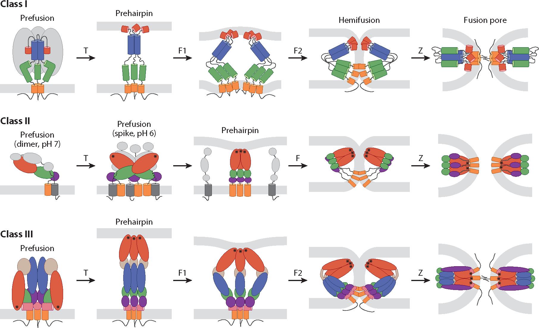
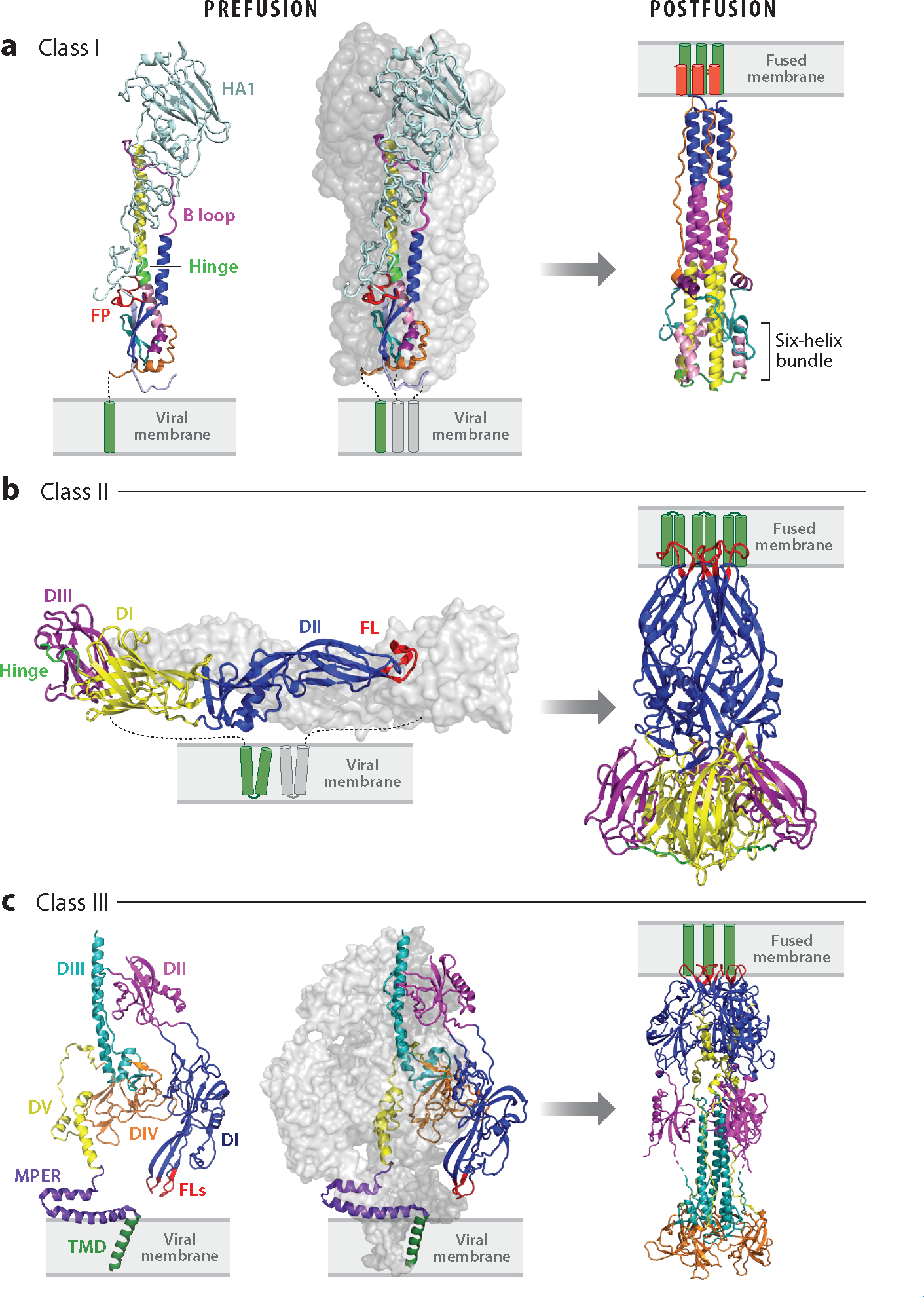
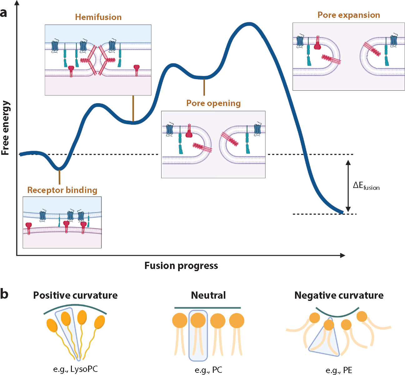
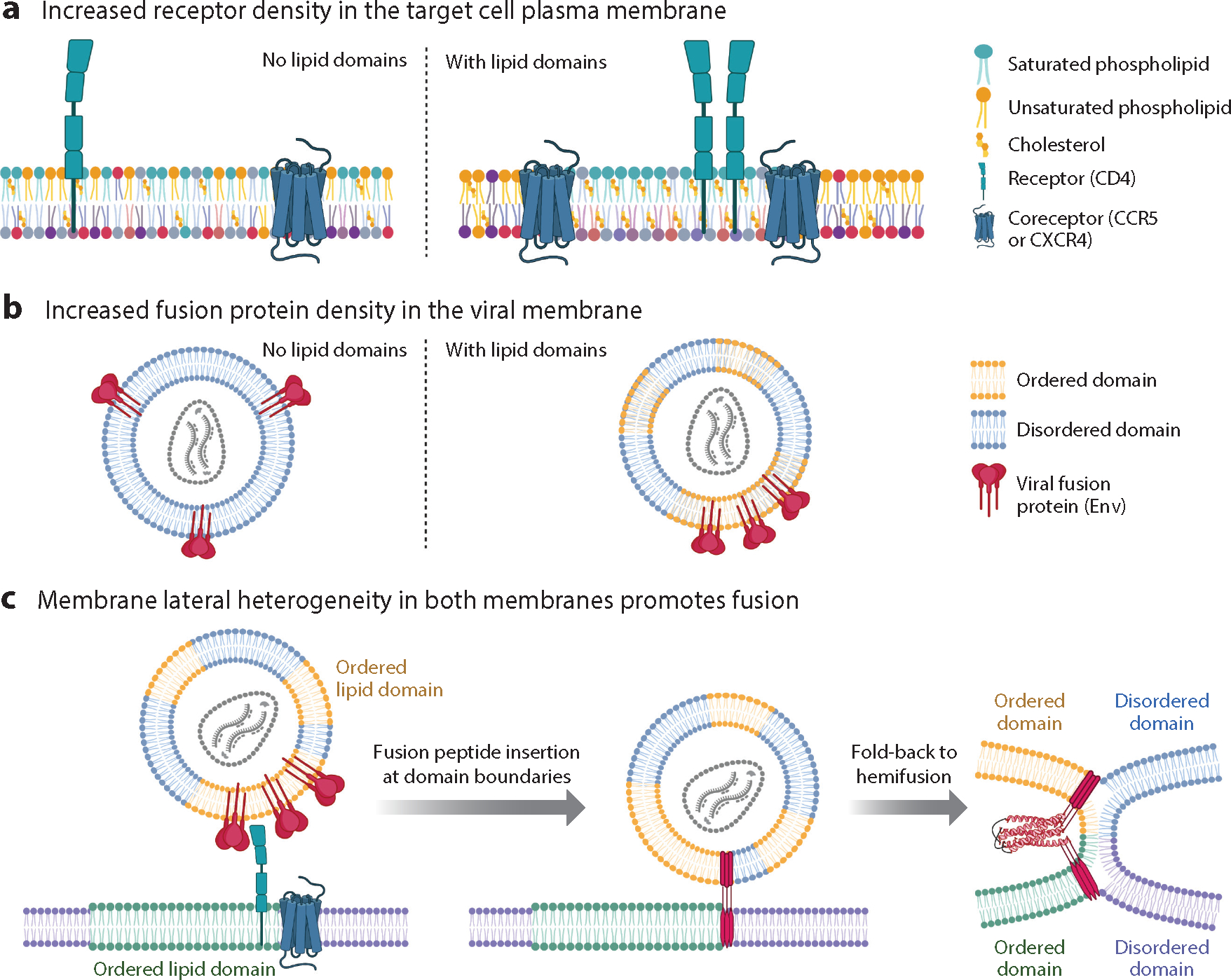
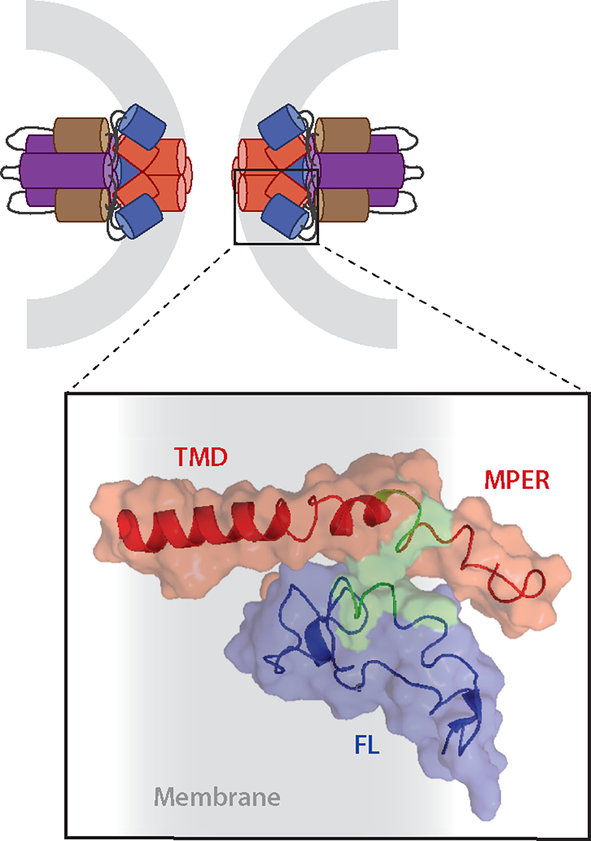
There are at least 21 families of enveloped viruses that infect mammals, and many contain members of high concern for global human health. All enveloped viruses have a dedicated fusion protein or fusion complex that enacts the critical genome-releasing membrane fusion event that is essential before viral replication within the host cell interior can begin. Because all enveloped viruses enter cells by fusion, it behooves us to know how viral fusion proteins function. Viral fusion proteins are also major targets of neutralizing antibodies, and hence they serve as key vaccine immunogens. Here we review current concepts about viral membrane fusion proteins focusing on how they are triggered, structural intermediates between pre- and postfusion forms, and their interplay with the lipid bilayers they engage. We also discuss cellular and therapeutic interventions that thwart virus-cell membrane fusion.

Keywords: class I viral fusion proteins; class II viral fusion proteins; class III viral fusion proteins; conformational intermediates; fusion energetics; fusion loops; fusion peptides; fusion restriction factors; lipid dynamics; trimers-of-hairpins.


Figures

Figure 1
Figure 1
Fusion pathways of class I, II, and III viral fusion proteins. The fusion proteins schematically depicted are human immunodeficiency virus (HIV) envelope (Env) (class I), Semliki Forest virus (SFV) E1 (class II), and herpes simplex virus 1 (HSV-1) gB (class III). In response to specific triggers (T), prefusion structures proceed through stages of extended prehairpin formation, fold-back (F), and zippering (Z) while the membranes progress from separated bilayers through hemifusion and fusion pore formation. F, F1, and F2 denote progressive stages of fold-back but are not meant to imply there are only one or two stages. Color coding and symbols are as follows: Class I: red, fusion peptide; blue, heptad repeat (HR) 1; green, HR2; orange, transmembrane domain (TMD); gray, HIV gp120. Class II, E1: green, domain I; red, domain II; purple, domain III; orange, TMD of E1; asterisks, fusion loops. Class II, (SFV) E2: gray, domains A, B, and C and TMD. Class III: red, domain I; light brown, domain II; blue, domain III; green, domain IV; purple, domain V; pink, membrane proximal external region (MPER); orange, TMD; asterisks, fusion loops. Thin black lines below TMDs denote cytoplasmic tails. See Supplemental Figure Legend 1 for more information and references.
Figure 2
Figure 2
Pre- and postfusion structures of class I, II, and III viral fusion proteins. (a) Class I: influenza hemagglutinin (HA) in pre- [Protein Data Bank (PDB) 2HMG] and postfusion (PDB 1QU1) states (far left, excised prefusion monomer). The ectodomains of two monomers of the trimer are shown as surface representations in shades of gray; the other is colored: light blue, HA1; red, fusion peptide (FP); blue, helix A; magenta, B loop prefusion and helix B postfusion; yellow, helix C; neon green, helix D prefusion and DE turn postfusion; pink, helix E; teal, loop F; purple, helix G; orange, C-terminal leash. Transmembrane domains (TMDs) are shown in green. HA1 is not seen in the postfusion structure. (b) Class II: dengue virus E in pre- (left, PDB 4UTB, side view) and postfusion (right, PDB 1OK8) conformations. In the left panel (prefusion), one E ectodomain monomer is shown in gray and the other is coded with domains I, II, and III in yellow, blue, and purple, respectively; the fusion loops (FLs) are shown in red, and the E TMDs are depicted in green. The companion protein, precursor membrane (prM), is not shown. All class II fusion proteins, including those involved in eukaryotic and archaeal fusion (187, 188), have the same basic architecture. (c) Class III: human cytomegalovirus (HCMV) gB is shown in pre- (PDB 7KDP, left two panels) and postfusion (PDB 7KDD) conformations; an excised monomer is shown on the far left. Domains I, II, III, IV, and V of one monomer are shown in blue, magenta, teal, orange, and yellow, respectively. The membrane proximal external region (MPER) is in purple, the TMD in green, and the FLs in red. Cytoplasmic tails are not shown in any panels. See Supplemental Figure Legend 2 for more information and additional references.
Figure 3
Figure 3
Energetics and membrane dynamics during fusion. (a) An approximated schema of fusion energetics as exemplified by the human immunodeficiency virus (HIV) envelope (Env) glycoprotein (protein symbols as in Figure 4). The blue curve represents approximate free energy from receptor binding to hemifusion to pore opening to pore expansion. The energies depicted are approximate, but the transition energies (peaks) range from ~10 to 100 kBT. (b) Lipids can have positive [lysophosphatidylcholine (lysoPC)], negative [phosphatidylethanolamine (PE)], or no [phosphatidylcholine (PC)] intrinsic membrane curvature. Negative intrinsic curvature of the exterior leaflet stabilizes hemifusion intermediates, and positive intrinsic curvature in the interior leaflets stabilizes fusion pores. Figure adapted from Reference with permission. See Supplemental Figure Legend 3 for more information and references.
Figure 4
Figure 4
Effects of membrane lateral heterogeneity on viral membrane fusion as exemplified by human immunodeficiency virus (HIV). (a) In target cell membranes, lipid nanodomains can organize and concentrate receptors and/or fusion triggers. For HIV, the receptor, CD4, partitions to ordered nanodomains [rich in saturated (teal) phospholipids and cholesterol] while the HIV coreceptor, CCR5 (fusion trigger), partitions to the domain boundary. (b) In the viral membrane, lipid nanodomains influence the spacing of fusion proteins. On immature HIV particles, envelope (Env) is relatively immobile. Upon maturation via proteolytic cleavage of the juxtamembrane Gag polyprotein, Env diffuses more rapidly to form clusters that facilitate fusion. Env has multiple sequences that promote association with ordered nanodomains (yellow phospholipids; cholesterol not depicted) including a cholesterol recognition amino acid consensus motif within the membrane proximal external region (MPER) and palmitoylation sites, and an additional cholesterol interacting domain in the cytoplasmic tail of gp41. Env partitioning to ordered domains might be cell type and HIV strain dependent. (c) Lateral heterogeneity affects the energetics of fusion. The fusion peptides in the gp41 prehairpin preferentially insert at discontinuities in bilayer thickness between ordered and disordered lipid nanodomains. Upon fusion, the joining of two ordered domains produces one larger domain with a lower ratio of perimeter/area than the starting smaller domains. This minimizes line tension at the domain boundary and contributes favorably to the energetics of fusion. Figure adapted from images created with BioRender.com. See Supplemental Figure Legend 4 for more information and references.
Figure 5
Figure 5
A complex between fusion peptides/fusion loops (FLs) and membrane proximal external region (MPER)-transmembrane domain (TMD) regions completes the fusion protein refolding process that accompanies fusion pore opening. (top) Illustration of the fusion pore stage of Ebola virus glycoprotein (GP) mediated fusion; only GP2 is shown, and it is color coded: purple and brown, respectively, the N- and C-terminal heptad repeats; blue, FL; red, MPER-TMD. Gray represents the recently merged membrane. (bottom) Blowup of the boxed region in which the nuclear magnetic resonance (NMR) structures in membrane mimetics of the Ebola virus FL (blue) and MPER-TMD (red) were docked based on experimental interaction constraints from fluorescence and NMR data. Green denotes interacting residues. Gray represents membrane. Figure adapted from Reference . See Supplemental Figure Legend 5 for more information and additional references.

References

    1. Mercer J, Lee JE, Saphire EO, Freeman SA. 2020. Snapshot: enveloped virus entry. Cell 182(3):786. - PMC - PubMed
    1. White JM, Delos SE, Brecher M, Schornberg K. 2008. Structures and mechanisms of viral membrane fusion proteins: multiple variations on a common theme. Crit. Rev. Biochem. Mol. Biol. 43(3):189–219 - PMC - PubMed
    1. Harrison SC. 2015. Viral membrane fusion. Virology 479–480:498–507 - PMC - PubMed
    1. White JM, Whittaker GR. 2016. Fusion of enveloped viruses in endosomes. Traffic 17(6):593–614 - PMC - PubMed
    1. Rey FA, Lok S-M. 2018. Common features of enveloped viruses and implications for immunogen design for next-generation vaccines. Cell 172(6):1319–34 - PMC - PubMed

Publication types

LinkOut - more resources