. 2023 Nov;32(11):e4792.
doi: 10.1002/pro.4792.

UCSF ChimeraX: Tools for structure building and analysis

Affiliations

UCSF ChimeraX: Tools for structure building and analysis

Elaine C Meng et al. Protein Sci. 2023 Nov.

Abstract

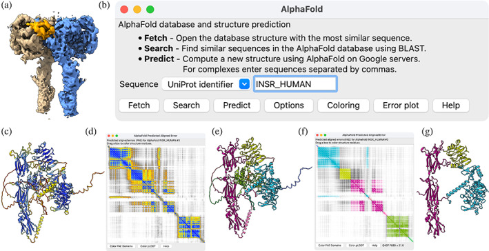
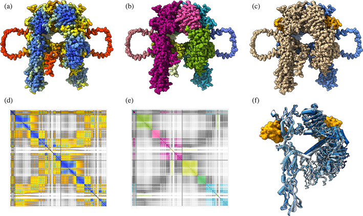
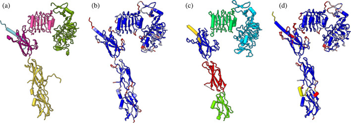
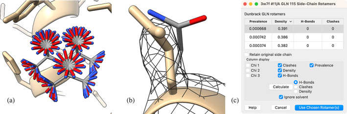
Advances in computational tools for atomic model building are leading to accurate models of large molecular assemblies seen in electron microscopy, often at challenging resolutions of 3-4 Å. We describe new methods in the UCSF ChimeraX molecular modeling package that take advantage of machine-learning structure predictions, provide likelihood-based fitting in maps, and compute per-residue scores to identify modeling errors. Additional model-building tools assist analysis of mutations, post-translational modifications, and interactions with ligands. We present the latest ChimeraX model-building capabilities, including several community-developed extensions. ChimeraX is available free of charge for noncommercial use at https://www.rbvi.ucsf.edu/chimerax.

Keywords: AlphaFold; ChimeraX; atomic model building; cryo-electron microscopy; protein structure prediction; refinement.


Conflict of interest statement

The authors declare no conflicts of interest.

Figures

FIGURE 1
FIGURE 1
AlphaFold Database prediction of the human insulin receptor. (a) cryo‐EM map of insulin receptor dimer (EMDB 34281; Kim et al., , 4.2 Å resolution) with insulin receptor monomers in brown and blue and insulin in orange. (b) ChimeraX AlphaFold tool user interface. (c) Human insulin receptor monomer prediction from the AlphaFold Database colored by pLDDT from blue (high confidence) to yellow to red (low confidence) does not match the experimental confirmation. (d) Predicted aligned error (PAE) plot with blue representing small errors (<5 Å), and yellow (10 Å), orange (15 Å), and gray (> 20 Å) representing larger estimated errors. (e) Structure colored by PAE domain, green cytoplasmic, pink transmembrane, purple fibronectin, and yellow and blue extracellular. (f) PAE plot colored by PAE domain. (g) Structure as in (e) except with the disordered and unobserved (in cryo‐EM, see text) parts removed.
FIGURE 2
FIGURE 2
AlphaFold prediction of dimeric insulin receptor. (a) AlphaFold 2.3 prediction of dimeric human insulin receptor with two insulin molecules, colored: by pLDDT score; (b) by PAE domains; (c) with receptor chains tan and blue, insulin orange. (d) PAE plot colored by PAE. (e) PAE plot colored by PAE domain. (f) Superposition of five predictions showing only one monomer. All five predictions are similar to one another.
FIGURE 3
FIGURE 3
AlphaFold and ESMFold domains aligned to cryo‐EM final structure. (a) Insulin receptor domains from AlphaFold prediction individually aligned to final cryo‐EM structure (PDB 8guy, not shown) for one insulin receptor monomer. (b) Final cryo‐EM structure colored by C‐alpha distance from AlphaFold domains (blue = 1 Å, white = 2 Å, red = 3 Å). (c) ESMFold individually predicted domains aligned to cryo‐EM structure. (d) Final cryo‐EM structure colored by C‐alpha distance from ESMFold domains.
FIGURE 4
FIGURE 4
AlphaFold predicts an insulin binding site not observed in the cryo‐EM map of the human structure. (a) Contacts <5 Å between insulin and receptor third fibronectin domain in the AlphaFold prediction shown as colored lines (dark blue: PAE 0, light blue: PAE 5, yellow: PAE 10) have high confidence. (b) AlphaFold dimer prediction with insulin bound to the side of the third fibronectin domain. (c) Final cryo‐EM structure (PDB 8guy) with insulin bound to top of third fibronectin domain. (d) Structure of mouse insulin receptor with two insulin binding locations (PDB 7sl7), the upper binding location matching the human cryo‐EM data. (e) Superposition of AlphaFold prediction (orange and tan) with mouse structure (yellow and pink) of lower insulin binding location.
FIGURE 5
FIGURE 5
Insulin and the five PAE domains of the AlphaFold‐predicted insulin receptor monomer fitted separately into the map to assemble an initial model.
FIGURE 6
FIGURE 6
Assessing model fit to map. (a) Atomic model surface depiction of topoisomerase (blue) bound to DNA (green and brown) with cyanotriazole inhibitor (yellow), PDB 8gcc. (b) Thin slab of 2.94 Å map (EMD 29930) showing atomic model side chains. (c) Sharpened cryo‐EM map colored by local resolution calculated by Phenix local_resolution (Anon Creating a local resolution map with local_resolution, n.d.). (d) QScore ChimeraX extension. (e) Strudel‐ Score ChimeraX extension.
FIGURE 7
FIGURE 7
Spatial arrangement of ribosomes seen in untreated cells and cells treated with anticancer drug homoharringtonine, with colors indicating the ribosome conformational state (Figure 4 of Xing et al., 2023). Ribosomes are more dispersed in drug‐treated cells.
FIGURE 8
FIGURE 8
B‐form DNA generated by ChimeraX drawn with simplified nucleotide representations and colored by residue type: A, red; C, yellow; G, green; T, blue.
FIGURE 9
FIGURE 9
(a) Rotamers from the Dunbrack backbone‐dependent rotamer library (Shapovalov & Dunbrack Jr, 2011) for glutamine 115 in chain A of dehydrosqualene synthase (PDB 3w7f). (b) Single rotamer with highest correlation to experimental density and no clashes (thin, element‐colored) along with experimentally placed side chain (thick, monocolored). (c) Rotamers table.

References

    1. Altschul SF, Gish W, Miller W, Myers EW, Lipman DJ. Basic local alignment search tool. J Mol Biol. 1990;215:403–410. - PubMed
    1. Anon AMBER parameter database . Available from: http://amber.manchester.ac.uk
    1. Anon Creating a local resolution map with local_resolution . Available from: https://phenix-online.org/documentation/reference/local_resolution.html
    1. Anon Google Colaboratory . Available from: https://colab.research.google.com/?utm_source=scs-index
    1. Bolton EE, Chen J, Kim S, Han L, He S, Shi W, et al. PubChem3D: a new resource for scientists. J Chem. 2011;3:32. - PMC - PubMed

Publication types