. 2023 Nov 30;142(22):1918-1927.
doi: 10.1182/blood.2022019282.

Cold exposure induces vaso-occlusion and pain in sickle mice that depend on complement activation

Affiliations

Cold exposure induces vaso-occlusion and pain in sickle mice that depend on complement activation

Zalaya K Ivy et al. Blood. .

Abstract

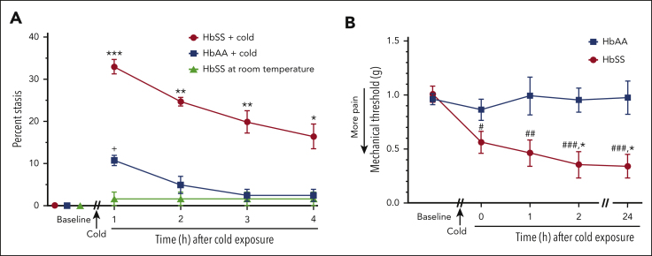
Vaso-occlusive pain episodes (VOE) cause severe pain in patients with sickle cell disease (SCD). Vaso-occlusive events promote ischemia/reperfusion pathobiology that activates complement. We hypothesized that complement activation is linked to VOE. We used cold to induce VOE in the Townes sickle homozygous for hemoglobin S (HbSS) mouse model and complement inhibitors to determine whether anaphylatoxin C5a mediates VOE. We used a dorsal skinfold chamber to measure microvascular stasis (vaso-occlusion) and von Frey filaments applied to the plantar surface of the hind paw to assess mechanical hyperalgesia in HbSS and control Townes mice homozygous for hemoglobin A (HbAA) mice after cold exposure at 10°C/50°F for 1 hour. Cold exposure induced more vaso-occlusion in nonhyperalgesic HbSS mice (33%) than in HbAA mice (11%) or HbSS mice left at room temperature (1%). Cold exposure also produced mechanical hyperalgesia as measured by paw withdrawal threshold in HbSS mice compared with that in HbAA mice or HbSS mice left at room temperature. Vaso-occlusion and hyperalgesia were associated with an increase in complement activation fragments Bb and C5a in plasma of HbSS mice after cold exposure. This was accompanied by an increase in proinflammatory NF-κB activation and VCAM-1 and ICAM-1 expression in the liver. Pretreatment of nonhyperalgesic HbSS mice before cold exposure with anti-C5 or anti-C5aR monoclonal antibodies (mAbs) decreased vaso-occlusion, mechanical hyperalgesia, complement activation, and liver inflammatory markers compared with pretreatment with control mAb. Anti-C5 or -C5aR mAb infusion also abrogated mechanical hyperalgesia in HbSS mice with ongoing hyperalgesia at baseline. These findings suggest that C5a promotes vaso-occlusion, pain, and inflammation during VOE and may play a role in chronic pain.


Conflict of interest statement

Conflict-of-interest disclosure: J. D. Belcher, G.M.V., and D.A.S. have research funding from Omeros. J. D. Belcher and G.M.V. have research funding from CSL Behring, Hillhurst Biopharmaceuticals, Sanofi, and Astellas/Mitobridge; and both are consultants for Sanofi and Astellas/Mitobridge. G.M.V. also is on data safety monitoring committees for Alexion and Novo Nordisk trials. K.G. reports honoraria from Novartis and CSL Behring and research grants from Cyclerion, 1910 Genetics, Novartis, Grifols, Zilker, UCI Foundation, and SCIRE Foundation. J. D. Beckman has research funding from Bayer. The remaining authors declare no competing financial interests.

Figures

None
Graphical abstract
Figure 1.
Figure 1.
Cold exposure induces vaso-occlusion and mechanical hyperalgesia in HbSS mice. (A) Townes HbSS and HbAA mice (n = 4 per group) were implanted with DSFC windows. At baseline, flowing subcutaneous venules (20-23 venules per mouse) were selected and mapped. HbSS and HbAA mice were exposed to cold at 10°C (50°F) for 1 hour and then returned to RT. A control group of HbSS mice remained at RT without cold exposure. Microvascular stasis (vaso-occlusion) was measured at the indicated times after cold exposure. Values are means ± standard error of the mean (SEM). ∗P < .05, ∗∗P < .01, and ∗∗∗P < .001, HbSS + cold vs HbAA + cold and HbSS at RT and +P < .05 HbAA + cold vs HbSS at RT (2-way analysis of variance [ANOVA] with the Tukey multiple comparison test). (B) Mechanical PWT was measured in the hind paws of HbAA and HbSS mice using von Frey filaments at baseline at RT. Nonhyperalgesic (PWT > 0.7 g) HbAA (n = 11) and HbSS (n = 14 mice) mice were selected for cold exposure. After baseline measurements, nonhyperalgesic mice were exposed to cold (10°C/50°F) for 1 hour and returned to RT. PWT was measured at the indicated times after cold exposure. Values are means ± SEM. ∗P < .05 HbSS vs HbAA (2-way ANOVA with the Sidak multiple comparison test). #P < .05, ##P < .01, and ####P < .0001, baseline vs after cold (2-way ANOVA with the Dunnett multiple comparison test).
Figure 2.
Figure 2.
Activated complement Bb and C5a fragments are increased in the plasma of HbSS mice after cold exposure. HbAA (n = 2 or 3 per group) and HbSS mice (n = 4 per group) were exposed to cold at 10°C (50°F) for 1 hour and then returned to RT or left untreated at RT. Four hours after cold exposure, mice were euthanized and EDTA blood samples were collected from the heart. EDTA plasma was run on immunoblots and stained for complement activation fragments (A) Bb, (B) C5a, and immunoglobulin G (IgG) loading controls. Zymosan-treated mouse serum, zymosan-treated heat-inactivated serum, and untreated mouse serum were analyzed on the C5a immunoblots to serve as positive and negative controls. Bb, C5a, and IgG bands on the immunoblot images were quantified using densitometry. Relative plasma Bb (C) and C5a (D) levels are presented as densities relative to IgG light chain control. Bars are means ± SEM. ∗∗P < .01 (1-way ANOVA with the Tukey multiple comparison test).
Figure 3.
Figure 3.
Vaso-occlusion and hyperalgesia induced by cold exposure are ameliorated by blocking C5 activation or C5aR signaling. (A) HbSS mice (n = 4 per group) were implanted with DSFC windows. At baseline, flowing subcutaneous venules (20-23 venules per mouse) were selected and mapped. After baseline selection of flowing venules, mice were injected IV via the tail vein with control, anti-C5 mAb, or anti-C5aR mAb (1.2 mg/kg). Thirty minutes after mAb infusion, mice were exposed to cold at 10°C (50°F) for 1 hour and then returned to RT. Microvascular stasis (vaso-occlusion) was measured at the indicated times after return to RT. Values are means ± SEM. ∗∗P < .01 and ∗P < .05, anti-C5 or anti-C5aR mAb vs control mAb (2-way ANOVA with the Tukey multiple comparison test). (B) Baseline PWT was measured in the hind paws of HbSS mice using von Frey filaments. After baseline pain measurements, nonhyperalgesic mice (PWT > 0.7) were injected IV via the tail vein with control mAb, anti-C5 mAb, or anti-C5aR mAb (n = 4 mice per group; 1.2 mg/kg). Thirty minutes after mAb infusion, mice were exposed to cold at 10°C (50°F) for 1 hour and then returned to RT. PWT was measured at the indicated times after cold exposure. (C) In experiments without cold exposure, HbSS mice that had ongoing hyperalgesia (PWT < 0.7) during baseline testing were injected IV via the tail vein with control mAb or anti-C5aR mAb (n = 6 per group; 1.2 mg/kg). PWT was measured at the indicated times after mAb infusion. (B-C) Values are means ± SEM. ∗P < .05 control mAb vs anti-C5 or anti-C5aR mAbs (2-way ANOVA with the Tukey multiple comparison test). #P < .05, ##P < .01, and ###P < .001 vs baseline (2-way ANOVA, with the Dunnett multiple comparison test).
Figure 4.
Figure 4.
Activated complement Bb and C5a fragments in plasma are decreased by blocking C5 activation or C5aR signaling. (A-B) After baseline pain testing, nonhyperalgesic HbSS mice were injected IV via the tail vein with control, anti-C5, or anti-C5aR mAbs (n= 4 per group; 1.2 mg/kg). Thirty minutes after mAb infusion, nonhyperalgesic mice were exposed to cold at 10°C (50°F) for 1 hour and then returned to RT. Four hours after mice were returned to RT, mice were euthanized and EDTA blood samples were collected from the heart. EDTA plasma samples were analyzed by immunoblots stained with antibodies to (A) Bb or (B) C5a activation fragments, and IgG loading controls. Zymosan-treated mouse serum, zymosan-treated heat-inactivated serum, and untreated mouse serum were analyzed on the C5a immunoblots to serve as positive and negative controls. (C) In experiments without cold exposure, HbSS mice that had ongoing hyperalgesia (PWT < 0.7) during baseline testing were injected IV via the tail vein with control or anti-C5aR mAbs (n = 4 per group; 1.2 mg/kg). EDTA blood samples were collected from the heart 4 hours after mAb infusion. EDTA plasma samples were analyzed by immunoblots stained with antibodies to Bb, C5a, and IgG. (D-G) The intensities of the Bb, C5a, and IgG light chain bands on the immunoblot images were measured using densitometry. Bb to IgG ratios in panel A are presented in panel D; C5a to IgG ratios in panel B are presented in panel E; Bb to IgG and C5a to IgG ratios in panel C are presented in panels F-G. Bars are means ± SEM. ∗P < .05, ∗∗P < .01, ∗∗∗P < .001, and ∗∗∗∗P < .0001 (1-way ANOVA with the Tukey multiple comparison test).
Figure 5.
Figure 5.
Vaso-occlusion and mechanical hyperalgesia induced by cold exposure are inhibited by anti-P-selectin mAb. (A) Anti–P-selectin mAb inhibited cold-induced vaso-occlusion in HbSS mice. Townes HbSS mice (n = 4 per group) were implanted with DSFC windows. After baseline selection of flowing subcutaneous venules, mice were injected IV via the tail vein with isotype control or anti–P-selectin mAb (1.2 mg/kg). Thirty minutes later, mice were exposed to cold at 10°C (50°F) for 1 hour and returned to RT. Microvascular stasis was measured in the same venules at the indicated times after cold exposure. Values are means ± SEM. ∗∗P < .01 and ∗∗∗P < .001, anti–P-selectin mAb vs control mAb (2-way ANOVA with the Sidak multiple comparison test). (B) Anti–P-selectin mAb inhibited cold-induced mechanical hyperalgesia in Townes HbSS mice. Baseline mechanical threshold was measured in the hind paws of HbSS mice (n = 4 mice per group) using von Frey filaments. After baseline pain measurements, nonhyperalgesic mice were injected IV via the tail vein with isotype control or anti–P-selectin mAb (1.2 mg/kg). Thirty minutes after mAb infusion, mice were exposed to cold at 10°C (50°F) for 1 hour and then returned to RT, and mechanical threshold was measured at the indicated times after cold exposure. (C) Anti–P-selectin mAb did not inhibit mechanical hyperalgesia in a complete Freund's adjuvant (CFA) pain model in nonsickle C57BL6 mice. Baseline mechanical threshold was measured in the hind paws of C57BL6 mice (n = 4 mice per group) using von Frey filaments. After baseline measurements, mice received a single injection of 10 μL of CFA into the middle of the plantar surface of 1 hind paw. Twenty-four hours after CFA injection, mice were injected IV via the tail vein with isotype control or anti–P-selectin mAb (1.2 mg/kg). Mechanical threshold was determined at the indicated times after mAb infusion. (B-C) Values are means ± SEM. ∗P < .05, anti–P-selectin mAb vs control mAb (2-way ANOVA with the Tukey multiple comparison test). #P < .05 and ##P < .01, vs baseline (2-way ANOVA, with the Dunnett multiple comparison test).
Figure 6.
Figure 6.
A circular model of cold-evoked vaso-occlusive pain crisis in SCD. These data are consistent with a feedback loop pain model whereby the outputs of the model, for example, C5a, are used as inputs to drive more inflammation, vaso-occlusion, and pain. In this model, cold exposure induces pain, vasoconstriction, and hypoxia,, likely leading to hemoglobin S (HbS) deoxygenation/polymerization, and microvascular stasis (vaso-occlusion). Static venules can subsequently reopen producing I/R pathophysiology leading to complement activation, including the cleavage of C5 and the generation of the potent anaphylatoxin C5a. Cleavage of C5 can be blocked by anti-C5 mAb. In the absence of anti-C5 mAb, the C5a produced can bind to C5aR and promote vascular inflammation and pain. The binding of C5a to C5aR and its subsequent proinflammatory signaling can be blocked by anti-C5aR mAb. In the absence of anti-C5aR mAb, vascular inflammation promotes the expression of adhesion molecules leading to more vaso-occlusion, which can be blocked by anti-adhesion therapies such as anti–P-selectin. Inflammatory pain mediators can sensitize nociceptors in tissues leading to pain.

Comment in

  • Cold comfort in sickle cell disease.
    Sparkenbaugh E, Little J. Sparkenbaugh E, et al. Blood. 2023 Nov 30;142(22):1854-1856. doi: 10.1182/blood.2023022621. Blood. 2023. PMID: 38032675 No abstract available.

References

    1. Platt OS, Thorington BD, Brambilla DJ, et al. Pain in sickle cell disease. Rates and risk factors. N Engl J Med. 1991;325(1):11–16. - PubMed
    1. Sagi V, Song-Naba WL, Benson BA, Joshi SS, Gupta K. Mouse models of pain in sickle cell disease. Curr Protoc Neurosci. 2018;85(1):e54. - PubMed
    1. Hebbel RP, Belcher JD, Vercellotti GM. The multifaceted role of ischemia/reperfusion in sickle cell anemia. J Clin Invest. 2020;130(3):1062–1072. - PMC - PubMed
    1. Kalambur VS, Mahaseth H, Bischof JC, et al. Microvascular blood flow and stasis in transgenic sickle mice: utility of a dorsal skin fold chamber for intravital microscopy. Am J Hematol. 2004;77(2):117–125. - PubMed
    1. Cain DM, Vang D, Simone DA, Hebbel RP, Gupta K. Mouse models for studying pain in sickle disease: effects of strain, age, and acuteness. Br J Haematol. 2012;156(4):535–544. - PMC - PubMed

Publication types