. 2023 Oct 10;18(10):1954-1971.
doi: 10.1016/j.stemcr.2023.08.014. Epub 2023 Sep 28.

Highly contractile 3D tissue engineered skeletal muscles from human iPSCs reveal similarities with primary myoblast-derived tissues

Affiliations

Highly contractile 3D tissue engineered skeletal muscles from human iPSCs reveal similarities with primary myoblast-derived tissues

Erik van der Wal et al. Stem Cell Reports. .

Abstract

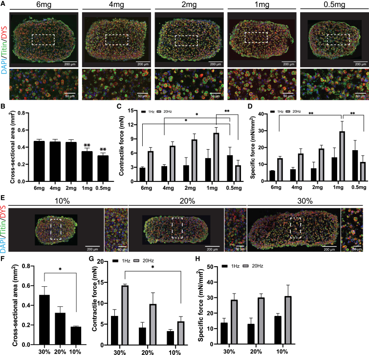
Skeletal muscle research is transitioning toward 3D tissue engineered in vitro models reproducing muscle's native architecture and supporting measurement of functionality. Human induced pluripotent stem cells (hiPSCs) offer high yields of cells for differentiation. It has been difficult to differentiate high-quality, pure 3D muscle tissues from hiPSCs that show contractile properties comparable to primary myoblast-derived tissues. Here, we present a transgene-free method for the generation of purified, expandable myogenic progenitors (MPs) from hiPSCs grown under feeder-free conditions. We defined a protocol with optimal hydrogel and medium conditions that allowed production of highly contractile 3D tissue engineered skeletal muscles with forces similar to primary myoblast-derived tissues. Gene expression and proteomic analysis between hiPSC-derived and primary myoblast-derived 3D tissues revealed a similar expression profile of proteins involved in myogenic differentiation and sarcomere function. The protocol should be generally applicable for the study of personalized human skeletal muscle tissue in health and disease.

Keywords: 3D-tissue engineering; contractile force; drug screening; induced pluripotent stem cells; myoblasts; myofiber; organ-on-a-chip; personalized medicine; satellite cell; skeletal muscle.


Conflict of interest statement

Declaration of interests A.I., E.W., V.S., W.P., and J.G. are inventors on a patent in the field of muscle on a chip.

Figures

Figure 1
Figure 1
Generation of myogenic progenitors under feeder-free conditions (A) Overview of the differentiation procedure. Top: feeder-based protocol as published previously (van der Wal et al., 2017). Bottom: feeder-free approach. (B) Expression of the early mesoderm marker Brachyury (green) and muscle stem cell marker PAX7 (red) analyzed by staining after 2 or 31 days of differentiation using the feeder-free differentiation protocol. Nuclei are stained with Hoechst. (C) Percentages of c-MET+/HNK fraction after 31 days of differentiation showing average ± standard deviation (SD) of 23 independent differentiations. (D) Average proliferation curve of myogenic progenitors from controls 1–3 during expansion culture. R2 was calculated from all data points. (E) Average cell cycle ±SD of control 1–3 myogenic progenitors during expansion culture derived from (D). (F) Staining of MYH, PAX7 or Hoechst after 4 days of differentiation of control 1–3 myogenic progenitors. Fusion index is shown in bottom corner as average percentage of nuclei inside MYH-positive cells ±SD and was quantified from five random fields.
Figure 2
Figure 2
Engineering of 3D-TESMs (A) Left: Schematic overview of experimental procedure and cartoon illustrating hydrogel compaction in Direct Peeling platform. Right: Overview of four culture conditions used. (B) 3D-TESMs in the Direct Peeling platform were cultured using conditions 1–4 and stained for titin (green) with whole-mount staining on day 7 of differentiation. Nuclei were visualized with DAPI (blue). (C) Immunofluorescent staining of cross-sections of 3D-TESMs visualized in (B) with antibodies against titin (green) and dystrophin (red), and counterstained with DAPI (blue).
Figure 3
Figure 3
Functional analysis of 3D-TESMs (A) Cartoon showing video-based contractile force measurement using an Arduino coupled with electrodes for stimulation and high-speed video imaging for recording of pillar displacement. (B) Sideview of a representative pillar of the Direct Peeling platform with a 3D-TESM attached. Position of the 3D-TESM is used for force calculation. (C) Top view of the pillar before and during 20-Hz stimulation. (D) Graph plotting displacement of the pillar upon 1-Hz stimulation for conditions 1–4 on day 7 of differentiation. (E) As (D) but then for a 20-Hz stimulation. (F) Same as (D) and (E) but then average absolute contractile force ±SD from three independent 3D-TESMs. Black bars indicate 1-Hz stimulation and gray bars 20-Hz stimulation. (G) Specific force of same 3D-TESMs as in (F) but then corrected for the cross-sectional area. ∗∗p < 0.01 using one-way ANOVA with Tukey multiple testing correction.
Figure 4
Figure 4
Determination of optimal hydrogel concentration for 3D-TESM formation (A) Representative cross-sections of 3D-TESMs generated with 6 mg–0.5 mg/mL of fibrinogen in the Direct Peeling platform and analyzed on day 7 of differentiation. Sections were stained with titin (green) and dystrophin (red). Nuclei were visualized in blue by DAPI staining. (B) Average cross-sectional area of 3D-TESMs generated with different concentrations of fibrinogen. (C) Average absolute contractile force of 3D-TESMs after stimulation with 1 Hz (black bars) or 20 Hz (gray bars). (D) Specific force of 3D-TESMs as in (C) but then normalized for cross-sectional area from (B). (E) Same as (A) but then for 3D-TESMs containing 30%–10% of Matrigel; 1 mg/mL fibrinogen was used. (F) Average cross-sectional area of 3D-TESMs containing 30%–10% of Matrigel. (G) Same as (C) but then for Matrigel concentration in 3D-TESMs. (H) Same as (G) but then corrected for cross-sectional area. p < 0.05, ∗∗p < 0.01 using one-way ANOVA with Tukey multiple testing correction. Data are derived from three independent 3D-TESMs and expressed as mean ± SD.
Figure 5
Figure 5
Comparison of the Direct Peeling platform with the Ecoflex Replica platform for miniaturization of 3D-TESMs (A) Cartoons showing (i) Direct Peeling (left) and Ecoflex Replica platform (right) fabrication schemes, (ii) 3D rendering of Direct Peeling and Ecoflex Replica culture chambers. (B) Whole-mount staining for titin (green) and nuclei with DAPI (blue) of 3D-TESMs on differentiation day 7. (C) Cross-sectional staining of a representative 3D-TESM. Antibodies against titin (green) and dystrophin (red) were used combined with DAPI nuclear staining (blue) to visualize myofibers. (D) Average number of dystrophin+ myofibers in cross-sections normalized for cross-sectional area. (E) Myofiber diameter of n > 100 myofibers (per section) positive for titin/dystrophin measured from n = 3 biological replicas. (F) Average absolute contractile force of 3D-TESMs stimulated with 1 Hz (black bars) and 20 Hz (gray bars). (G) Same as (F) but then specific force (normalized for cross-sectional area). (H) Average absolute contractile force of 3D-TESMs on day 7 of differentiation in Ecoflex platform after 6 h of incubation with caffeine, chloroquine, or cardiotoxin (CTX). 3D-TESMs were stimulated with either 1 Hz (black bars) or 20 Hz (gray bars). (I) Same as (H) but then for 3D-TESMs treated for 1 h with verapamil. N.C. in (H) and (I) stands for not contractile. p < 0.05, ∗∗p < 0.01, ∗∗∗∗p < 0.0001 using either independent-samples t test (D–G) or one-way ANOVA with Tukey multiple testing correction (H). Data are derived from three (D–G) or six to 10 (H and I) independent 3D-TESMs and expressed as mean ± SD.
Figure 6
Figure 6
3D-TESM generation of multiple donors and comparison with 3D-TESMs generated from primary myoblasts (A) 3D-TESMs of control 1–3 myogenic progenitors and primary myoblasts in the Ecoflex platform were stained for titin (green) and nuclei were visualized with DAPI (blue) on day 7 of differentiation. (B) Titin (green) and dystrophin (red) labeling of cross-sections generated from control 1–3 3D-TESMs or 3D-TESMs from primary myoblasts line 1. Nuclei were stained with DAPI (blue). (C) Comparison of the number of dystrophin+ myofibers in 3D-TESMs from different lines. (D) Myofiber diameter of n > 120 myofibers (3–4 cross-sections) quantified from n = 3 tissues per line. (E) Average absolute contractile force after stimulation with 1 Hz (black bars) and 20 Hz (gray bars). (F) Same as (E), but then specific force. (G) Relative mRNA expression of MYH isoforms in 3D-TESMs from control 1–3 myogenic progenitors and primary myoblasts line 1. Data were normalized for GUSB expression. p < 0.05, ∗∗p < 0.01 using one-way ANOVA with Tukey (C) or Games-Howell multiple testing correction (E, F). Data are derived from three (C, D, and G) or three to six (E and F) independent 3D-TESMs and expressed as mean ± SD.
Figure 7
Figure 7
Proteomic analysis of hiPSC-derived and primary myoblast-derived 3D-TESMs (A) Difference in LFQ intensity (normalized by column-wise median subtraction for each sample) between day 0 and day 7 of main MYH isoforms commonly associated with skeletal muscle, detected in control 1–3 hiPSC-derived 3D-TESMs and primary myoblast-derived ones. (B) Volcano plot for all three control lines and primary 3D-TESMs, showing fold change of all proteins identified at day 7 compared with day 0, based on LFQ intensity. An additional subset of myogenic markers is highlighted in blue and shown in the bottom-right box. (C) Gene ontology enrichment analysis performed for controls 1–3 and primary 3D-TESMs. Comparisons were made between day 0 and day 7 of differentiation for each line separately. Top five significantly enriched pathways are shown for each respective control. (D) Heatmap showing normalized LFQ intensity of selected proteins with highest sample set to 100% for each respective protein. Proteins were selected based on their association with development and function of skeletal muscle tissue, for each line at day 0 and day 7 of differentiation. (E) Volcano plots comparing hiPSC-derived 3D-TESMs with primary myoblast-derived 3D-TESMs. Comparisons of iBAQ intensity of each protein, with a subset of myogenic markers highlighted in blue in the bottom-right box. Data are derived from three independent 3D-TESMs and expressed as mean ± SD or as mean.

References

    1. Afshar M.E., Abraha H.Y., Bakooshli M.A., Davoudi S., Thavandiran N., Tung K., Ahn H., Ginsberg H.J., Zandstra P.W., Gilbert P.M. A 96-well culture platform enables longitudinal analyses of engineered human skeletal muscle microtissue strength. Sci. Rep. 2020;10:6918. doi: 10.1038/s41598-020-62837-8. - DOI - PMC - PubMed
    1. Afshar Bakooshli M., Lippmann E.S., Mulcahy B., Iyer N., Nguyen C.T., Tung K., Stewart B.A., van den Dorpel H., Fuehrmann T., Shoichet M., et al. A 3D culture model of innervated human skeletal muscle enables studies of the adult neuromuscular junction. Elife. 2019;8 doi: 10.7554/eLife.44530. - DOI - PMC - PubMed
    1. Benarroch L., Bonne G., Rivier F., Hamroun D. The 2020 version of the gene table of neuromuscular disorders (nuclear genome) Neuromuscul. Disord. 2019;29:980–1018. doi: 10.1016/j.nmd.2019.10.010. - DOI - PubMed
    1. Bigot A., Jacquemin V., Debacq-Chainiaux F., Butler-Browne G.S., Toussaint O., Furling D., Mouly V. Replicative aging down-regulates the myogenic regulatory factors in human myoblasts. Biol. Cell. 2008;100:189–199. doi: 10.1042/bc20070085. - DOI - PubMed
    1. Borchin B., Chen J., Barberi T. Derivation and FACS-mediated purification of PAX3+/PAX7+ skeletal muscle precursors from human pluripotent stem cells. Stem Cell Rep. 2013;1:620–631. doi: 10.1016/j.stemcr.2013.10.007. - DOI - PMC - PubMed

LinkOut - more resources