. 2023 Dec;35(52):e2306765.
doi: 10.1002/adma.202306765. Epub 2023 Nov 22.

Facile Photopatterning of Perfusable Microchannels in Synthetic Hydrogels to Recreate Microphysiological Environments

Affiliations

Facile Photopatterning of Perfusable Microchannels in Synthetic Hydrogels to Recreate Microphysiological Environments

Ana Mora-Boza et al. Adv Mater. 2023 Dec.

Abstract

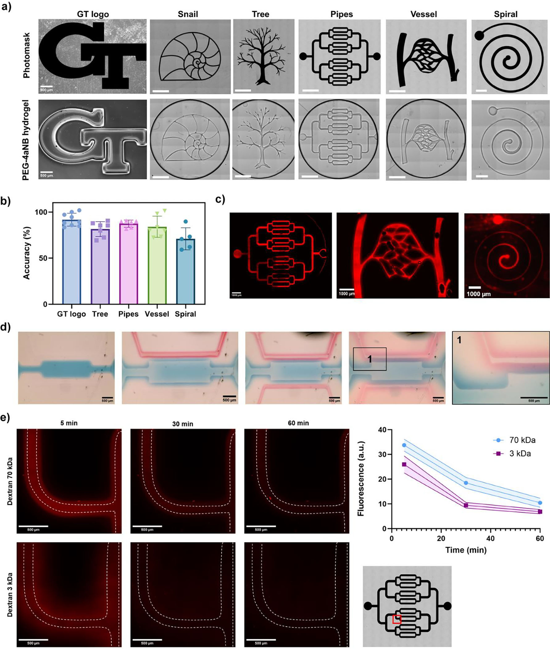
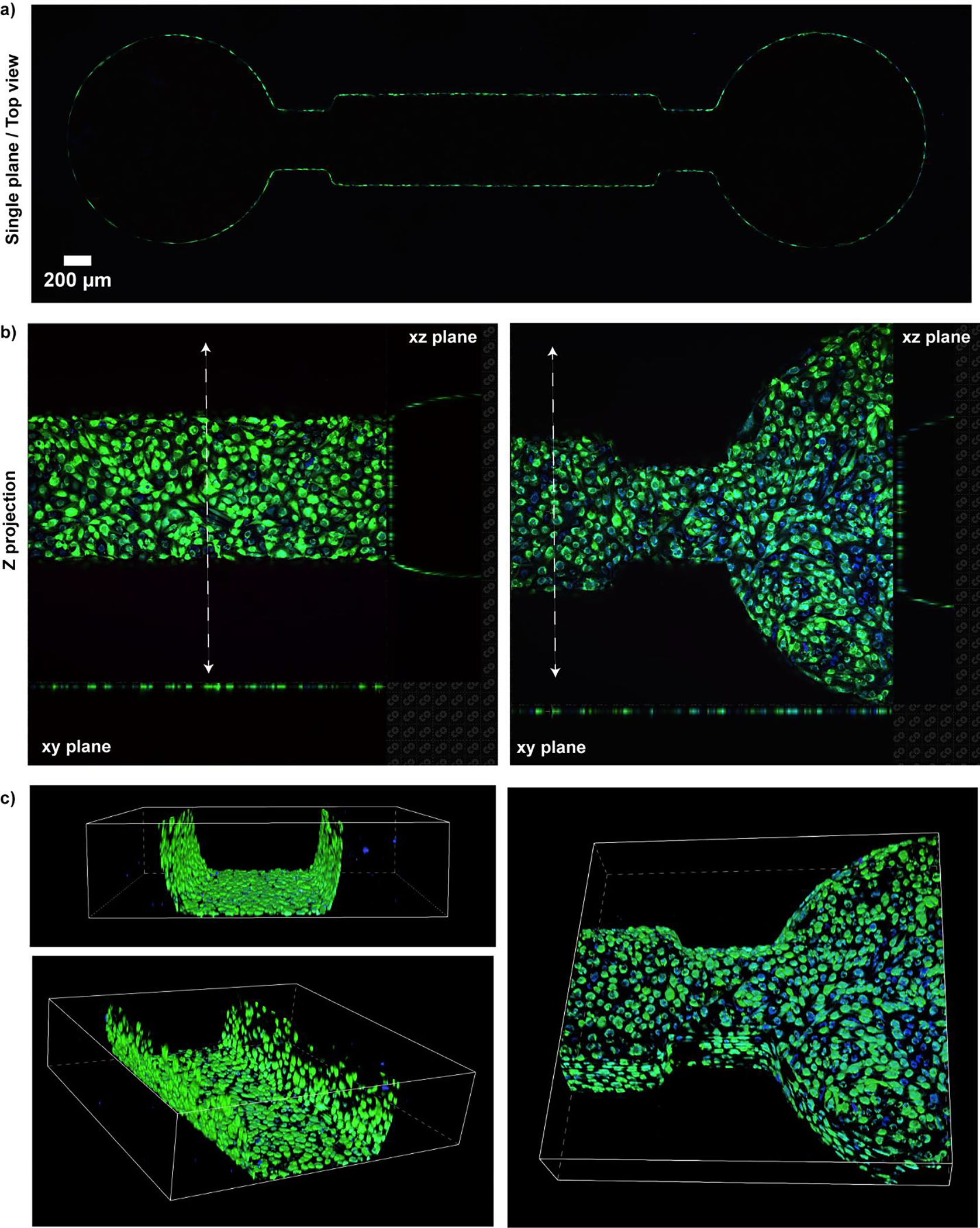
The fabrication of perfusable hydrogels is crucial for recreating in vitro microphysiological environments. Existing strategies to fabricate complex microchannels in hydrogels involve sophisticated equipment/techniques. A cost-effective, facile, versatile, and ultra-fast methodology is reported to fabricate perfusable microchannels of complex shapes in photopolymerizable hydrogels without the need of specialized equipment or sophisticated protocols. The methodology utilizes one-step ultraviolet (UV) light-triggered cross-linking and a photomask printed on inexpensive transparent films to photopattern PEG-norbornene hydrogels. Complex and intricate patterns with high resolution, including perfusable microchannels, can be fabricated in <1 s. The perfusable hydrogel is integrated into a custom-made microfluidic device that permits connection to external pump systems, allowing continuous fluid perfusion into the microchannels. Under dynamic culture, human endothelial cells form a functional and confluent endothelial monolayer that remains viable for at least 7 days and respond to inflammatory stimuli. Finally, approach to photopattern norbornene hyaluronic acid hydrogels is adapted, highlighting the versatility of the technique. This study presents an innovative strategy to simplify and reduce the cost of biofabrication techniques for developing functional in vitro models using perfusable three-dimensional (3D) hydrogels. The approach offers a novel solution to overcome the complexities associated with existing methods, allowing engineering advanced in vitro microphysiological environments.

Keywords: endothelial cells; hydrogels; microchannels; microfluidics; microphysiological systems; perfusion.


Figures

Figure 1:
Figure 1:
Photopatterning approach to fabricate in situ perfusable microchannels in PEG-4aNB hydrogels and hydrogel characterization. a) Hydrogel components comprise 4-arm amide-linked norbornene PEG (PEG-4aNB) macromer, RGD peptide (cell adhesive ligand), DTT or PEG-DT (crosslinker), and LAP (photoinitiator). The photopatterning strategy used for the fabrication of perfusable microchannels in hydrogels involves injecting PEG-4aNB solution into a microfluidic device (Figure S1) and applying UV light for in situ photopolymerization through a photomask. b) Swelling percentages for PEG-4aNB hydrogels fabricated with 5 or 20 kDa macromers at 10 and 12 wt%, using DTT or PEG-DT as crosslinkers. c) Storage and loss moduli of PEG-4aNB hydrogels fabricated with 5 or 20 kDa macromers at 10 and 12 wt%, using DTT or PEG-DT as crosslinkers. n = 5, *p<0.05, **p<0.005, ***p=0.0002, ****p<0.0001. Two-way ANOVA analysis was used to detect significant differences among groups.
Figure 2:
Figure 2:
Images of photopatterned designs in PEG-4aNB hydrogels, assessment of resolution, and demonstration of microchannel perfusability. a) Brightfield images of photomask transparencies used to generate photopatterns in PEG-4aNB hydrogels. Scale bar: 1 mm (unless indicated otherwise). b) Accuracy percentage (%) of photopatterned features in PEG-4aNB hydrogels for different photomasks; mean ± SD, n = 5–10 measurements per design. c) Fluorescence images displaying different photopatterned microchannels fabricated in PEG-4aNB hydrogels perfused with 70 kDa dextran molecule. d) Brightfield images of photopatterning of 3 independent microchannels in PEG-4aNB hydrogels. Scale bar: 500 μm. e) Fluorescence images of microchannels perfused with 70 and 3 kDa dextran molecules over time, and fluorescence intensity quantification of 70 and 3 kDa dextran molecules perfused over time (mean ± SD, n = 5). Photomask design is provided for reference. Red square indicates the region where the fluorescence images where captured.
Figure 3:
Figure 3:
Live/dead fluorescence imaging of cultured HUVECs in microchannels with different designs after 2 and 7 days of dynamic culture. a) Live/dead fluorescence images of HUVECs cultured in a single-channel device under perfusion for 2 and 7 days. Green represents GFP+ HUVECs; blue represents live cells stained with BioTracker 405 Blue dye; and red indicated dead cells stained with TOTO-3. Images labelled as 1 and 2 correspond to higher magnification images captured in the middle of the microchannels (1) and one of the outlets (2). b) GFP+ HUVECs cultured for 2 and 7 days in photopatterned multichannel design in PEG-4aNB hydrogels. All fluorescent images displayed are z-projections.
Figure 4:
Figure 4:
Live/dead fluorescence images of GFP+ HUVECs cultured in the devices for 7 days. a) Fluorescence image of a single plane of the device (top view) after 7 days of dynamic culture. b) Fluorescence images showing z-projections in different regions of a single-microchannel device, including projections for the xz and xy planes. c) 3D projection depicting different areas of the channel after 7 days of dynamic culture. GFP+ HUVECs are represented in green, while live cells stained with BioTracker 405 Blue dye are shown in blue.
Figure 5:
Figure 5:
Evaluation of barrier functionality and responsiveness of HUVECs cultured in single-microchannels devices with and without TNF-α exposure. a) Fluorescence images of immunostained HUVECs cultured with and without TNF-α. Green represents GFP+ HUVECs; blue represents nuclei stained with DAPI; and pink highlights CD31+ cells (i.e. endothelial tight junctions). Images labelled as 1 correspond to higher magnification images captured in the middle of the microchannels. b) Quantification of fluorescence intensities in different regions of the PEG-4aNB hydrogel matrix over time for untreated and treated samples with TNF-α. Mean ± SD, n = 3.
Figure 6:
Figure 6:
Images of photopatterned designs in NorHA hydrogels and assessment of resolutiona) Bright field images of photomask transparencies used to generate photopatterns in NorHA hydrogels. Scale bar: 1 mm. b) Accuracy percentage (%) of photopatterned features in NoRha hydrogels for different photomasks; mean ± SD, n = 5–10 measurements per design.

References

    1. Xue D, Wang Y, Zhang J, Mei D, Wang Y, Chen S, ACS Appl Mater Interfaces 2018, 10, 19428; - PubMed
    2. Heintz KA, Bregenzer ME, Mantle JL, Lee KH, West JL, Slater JH, Adv Healthc Mater 2016, 5, 2153. - PMC - PubMed
    1. Peters JT, Wechsler ME, Peppas NA, Regenerative Biomaterials 2021, 8; - PMC - PubMed
    2. Xie R, Zheng W, Guan L, Ai Y, Liang Q, Small 2020, 16, e1902838. - PubMed
    1. Menon NV, Tay HM, Wee SN, Li KHH, Hou HW, Lab Chip 2017, 17, 2960; - PubMed
    2. Chen R, Wang B, Liu Y, He J, Lin R, Li D, Biomed Eng Online 2019, 18, 87; - PMC - PubMed
    3. Madl CM, Heilshorn SC, Annual Review of Biomedical Engineering 2018, 20, 21; - PMC - PubMed
    4. Nichol JW, Koshy ST, Bae H, Hwang CM, Yamanlar S, Khademhosseini A, Biomaterials 2010, 31, 5536; - PMC - PubMed
    5. Ko J, Ahn J, Kim S, Lee Y, Lee J, Park D, Jeon NL, Lab Chip 2019, 19, 2822; - PubMed
    6. Zhang B, Lai BFL, Xie R, Davenport Huyer L, Montgomery M, Radisic M, Nat Protoc 2018, 13, 1793. - PubMed
    1. Tehranirokh M, Kouzani AZ, Francis PS, Kanwar JR, Biomicrofluidics 2013, 7, 51502; - PMC - PubMed
    2. Raj M K, Chakraborty S, Journal of Applied Polymer Science 2020, 137, 48958;
    3. Cameron TC, Randhawa A, Grist SM, Bennet T, Hua J, Alde LG, Caffrey TM, Wellington CL, Cheung KC, Micromachines 2022, 13, 1573. - PMC - PubMed
    1. Liu Z, Zhang Y, Yang T, Liu Y, Zhou W, Wang Z, Liu Y, Kong T, Journal of Materials Chemistry C 2020, 8, 2320.

LinkOut - more resources