. 2023 Sep;11(9):e007174.
doi: 10.1136/jitc-2023-007174.

Antitumor activity of the investigational B7-H3 antibody-drug conjugate, vobramitamab duocarmazine, in preclinical models of neuroblastoma

Affiliations

Antitumor activity of the investigational B7-H3 antibody-drug conjugate, vobramitamab duocarmazine, in preclinical models of neuroblastoma

Chiara Brignole et al. J Immunother Cancer. 2023 Sep.

Abstract

Introduction: B7-H3 is a potential target for pediatric cancers, including neuroblastoma (NB). Vobramitamab duocarmazine (also referred to as MGC018 and herein referred to as vobra duo) is an investigational duocarmycin-based antibody-drug conjugate (ADC) directed against the B7-H3 antigen. It is composed of an anti-B7-H3 humanized IgG1/kappa monoclonal antibody chemically conjugated through a cleavable valine-citrulline linker to a duocarmycin-hydroxybenzamide azaindole (vc-seco-DUBA). Vobra duo has shown preliminary clinical activity in B7-H3-expressing tumors.

Methods: B7-H3 expression was evaluated by flow-cytometry in a panel of human NB cell lines. Cytotoxicity was evaluated in monolayer and in multicellular tumor spheroid (MCTS) models by the water-soluble tetrazolium salt,MTS, proliferation assay and Cell Titer Glo 3D cell viability assay, respectively. Apoptotic cell death was investigated by annexin V staining. Orthotopic, pseudometastatic, and resected mouse NB models were developed to mimic disease conditions related to primary tumor growth, metastases, and circulating tumor cells with minimal residual disease, respectively.

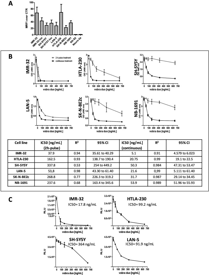
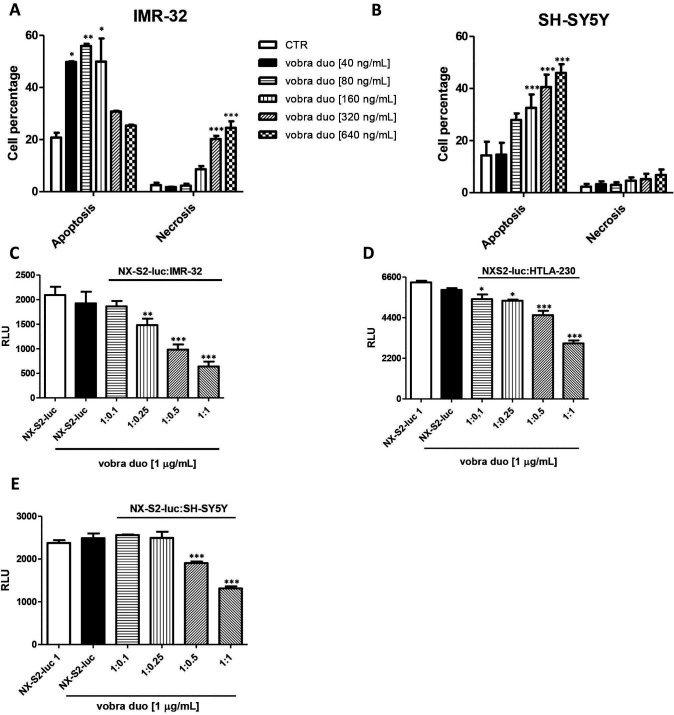
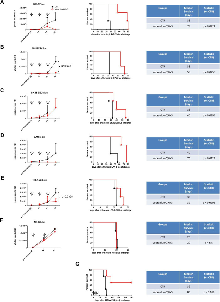
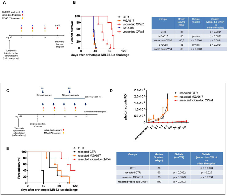
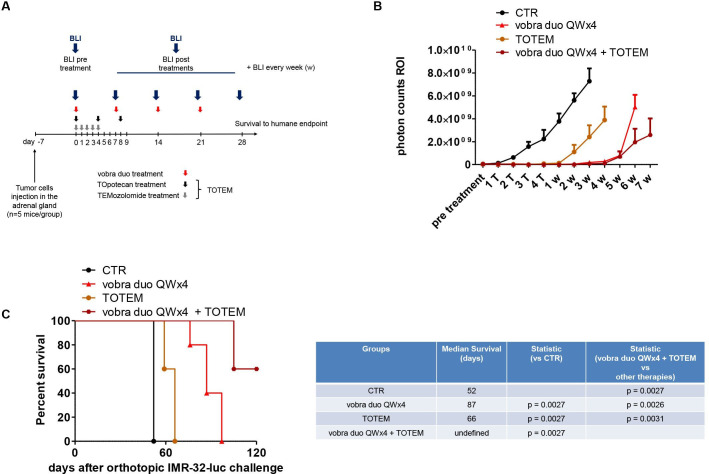
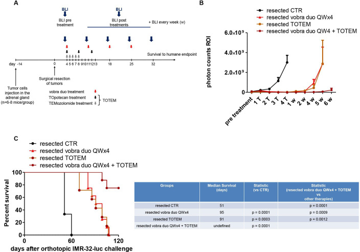
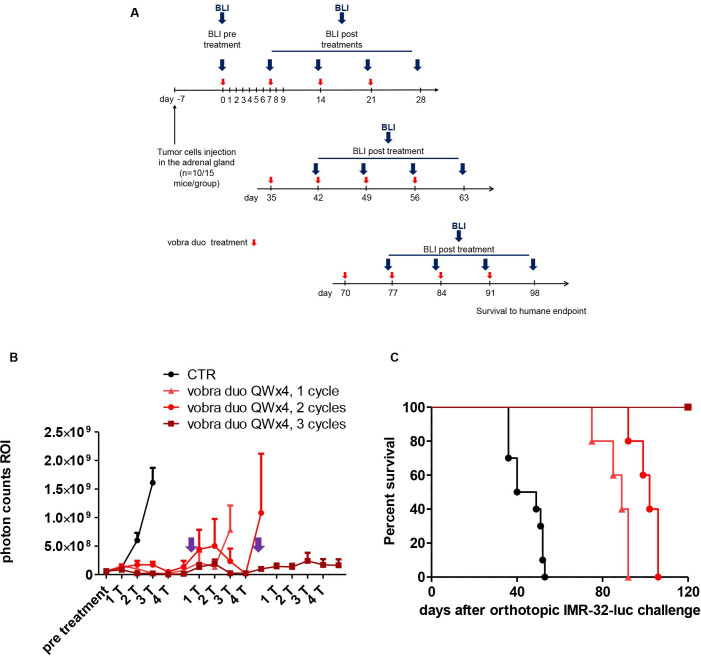
Results: All human NB cell lines expressed cell surface B7-H3 in a unimodal fashion. Vobra duo was cytotoxic in a dose-dependent and time-dependent manner against all cell lines (IC50 range 5.1-53.9 ng/mL) and NB MCTS (IC50 range 17.8-364 ng/mL). Vobra duo was inactive against a murine NB cell line (NX-S2) that did not express human B7-H3; however, NX-S2 cells were killed in the presence of vobra duo when co-cultured with human B7-H3-expressing cells, demonstrating bystander activity. In orthotopic and pseudometastatic mouse models, weekly intravenous treatments with 1 mg/kg vobra duo for 3 weeks delayed tumor growth compared with animals treated with an irrelevant (anti-CD20) duocarmycin-ADC. Vobra duo treatment for 4 weeks further increased survival in both orthotopic and resected NB models. Vobra duo compared favorably to TOpotecan-TEMozolomide (TOTEM), the standard-of-care therapy for NB relapsed disease, with tumor relapse delayed or arrested by two or three repeated 4-week vobra duo treatments, respectively. Further increased survival was observed in mice treated with vobra duo in combination with TOTEM. Vobra duo treatment was not associated with body weight loss, hematological toxicity, or clinical chemistry abnormalities.

Conclusion: Vobra duo exerts relevant antitumor activity in preclinical B7-H3-expressing NB models and represents a potential candidate for clinical translation.

Keywords: drug evaluation, preclinical; immunotherapy; neuroblastoma.


Conflict of interest statement

Competing interests: EB and DL are employees of MacroGenics and receive salary and stocks as part of their compensations. The other authors do not have competing interests to declare.

Figures

Figure 1
Figure 1
Cytotoxic activity of vobra duo against a panel of human B7-H3 positive NB cell lines. (A) The histograms represent the expression of human B7-H3 by human and murine NB cell lines. Protein expression was evaluated by flow cytometry and is expressed as mean ratio fluorescence intensity (MRFI) of positive labeled cells over cells stained with an isotype matched control Ab. (B) NB cell lines were treated with escalating concentrations of vobra duo (40-80-160-320-640 ng/mL) continuously for 7 days (black square) or for a 2-hour pulse followed by a washing step and continued incubation (gray circles). Viability was assessed by the MTS assay. Data are expressed as mean±SD. Vobra duo cytotoxicity IC50, calculated for both treatment schedules is reported. (C) Multicellular tumor spheroids models of four different NB cell lines were treated with increasing concentrations of vobra duo (40, 80, 160, 320, 640 ng/mL) for 7 days. Cell viability was determined by the Cell Titer Glo 3D Cell Viability Assay. Data are expressed as mean±SD. Ab, antibody; NB, neuroblastoma; MTS, water-soluble tetrazolium salt; OD, optical density; RLU, relative luminescence intensity; vobra duo, vobramitamab duocarmazine.
Figure 2
Figure 2
Vobra duo induces apoptotic and necrotic cell death and mediates bystander cell killing. (A, B) IMR-32 and SH-SY5Y cells were seeded in 6-well plates (4.5×10⁵ cells/well). The day after seeding, cells were treated with vobra duo for 24 hours, harvested and processed for the detection of apoptosis or necrosis. The percentage of apoptosis and necrosis induced by vobra duo is indicated as mean±SD. (C–E) Human B7-H3-negative NX-S2-luc cells (4×10³ cells/well) were co-cultured in black 96-well plates at different ratio (1:0.1, 1:0.25, 1:0.5 and 1:1) with IMR-32, HTLA-230, or SH-SY5Y cells and treated with vobra duo at the concentration of 1 µg/mL for 5 days. The graph represents the viability of NX-S2-luc cells expressed as relative luminescence unit (RLU, mean±SD). luc, luciferase; vobra duo, vobramitamab duocarmazine.
Figure 3
Figure 3
Antitumor effects of vobra duo in orthotopic and pseudometastatic murine models of NB. Mice, 7 days (A–F) or 4 hours (G) after being inoculated with NB cells, were randomly assigned to two treatment groups and intravenously treated with PBS (CTR) or 1 mg/kg vobra duo once a week for 3 weeks (vobra duo QWx3). (A–F) BLI curves and survival in luciferase (luc)-transfected human (A–E) and murine (F) orthotopic NB models (n=3 mice/group). For each cell line injected, tumor growth was monitored by BLI (left panels). Photon counts in the tumor region of interest (ROI) are reported at pre treatment and 7 days post treatment. X axes show the days after tumor cells injection. Results are presented as mean±SEM, with comparison of vobra duo versus CTR reported when statistically significant. Survival curves and statistical comparison of vobra duo QWx3 versus CTR are shown. (G) Survival in the pseudometastatic model (n=4 mice/group). Statistical comparison of vobra duo QWx3 versus CTR is reported. Arrow: treatment (T). BLI, bio-luminescence imaging; i.v., intravenous; NB, neuroblastoma; PBS, phosphate-buffered saline; QWx3, one time a week for 3 weeks; vobra duo, vobramitamab duocarmazine.
Figure 4
Figure 4
Vobra duo therapeutic efficacy in an orthotopic and in a primary tumor-resected model of NB. (A) Schematic and timeline of the experiment. IMR-32-luc cells were orthotopically inoculated in the mouse left adrenal gland. Treatments (MGA017, vobra duo QWx3, SYD988, vobra duo QWx4) started 7 days post cell inoculation and were performed as reported in the Results section. (B) Survival in NB-bearing mice treated with either vobra duo QWx3 (n=8 mice) or vobra duo QWx4 (n=5 mice). Statistics (vobra duo QWx3, MGA017 and SYD988 vs CTR; vobra duo QWx4 vs all groups) are reported. (C) Schematic and timelines of the experiment. IMR-32-luc cells were orthotopically inoculated in the mouse left adrenal gland. Tumors were surgically resected 2 weeks after, and treatments (n=5 mice/group) started 4 days post tumor resection, as reported. Untreated, control mice (CTR) received PBS. (D) Tumor growth was monitored by BLI. Photon counts in the tumor region of interest (ROI) are reported at pretreatment and 7 days post each treatment (T) and for the subsequent 4 weeks (w). Results are presented as mean±SEM. (E) Kaplan-Meier survival curves with corresponding statistical analysis comparing all therapeutic groups with vobra duo QWx4 are shown. CTR: mice orthotopically injected with tumor cells, and that did not undergo tumor surgical resection. BLI, bio-luminescence imaging; luc, luciferase; NB, neuroblastoma; QWx3, one time a week for 3 weeks; QWx4, one time a week for 4 weeks; vobra duo, vobramitamab duocarmazine.
Figure 5
Figure 5
Treatment combination of vobra duo and TOTEM in an orthotopic model of NB. (A) Schematic and timelines of the in vivo experiments. IMR-32-luc cells were orthotopically inoculated in the left adrenal gland of mice. Treatments (n=5 mice/group; vobra duo QWx4, TOTEM, vobra duo QWx4+TOTEM) started 7 days post cell inoculation and were performed as reported. Untreated, control mice (CTR) received PBS. (B) Tumor growth monitored by BLI. Photon counts in the tumor region of interest (ROI) are reported at pretreatment, 7 days post each treatment (T) and for the subsequent 7 weeks (w). Results are presented as mean±SEM. (C) Kaplan-Meier survival curves with corresponding statistical analysis comparing all therapeutic groups with vobra duo QWx4 are shown. BLI, bio-luminescence imaging; luc, luciferase; NB, neuroblastoma; PBS, phosphate-buffered saline; QWx4, one time a week for 4 weeks; vobra duo, vobramitamab duocarmazine.
Figure 6
Figure 6
Treatment combination of vobra duo and TOTEM in a primary tumor-resected model of NB. (A) Schematic and timeline of the in vivo experiments. IMR-32-luc cells were orthotopically inoculated in the left adrenal gland of mice. Tumors were surgically resected 2 weeks after, and treatments (n=5, CTR mice, n=8, treatments groups) started 4 days post tumor resection, as reported (vobra duo QWx4, TOTEM, vobra duo QWx4+TOTEM). Untreated, control mice (CTR) received PBS. (B) Tumor growth monitored by BLI. Photon counts in the tumor region of interest (ROI) are reported at pretreatment, 7 days post each treatment (T) and for the subsequent 6 weeks (w). Results are presented as mean±SEM. (C) Kaplan-Meier survival curves with corresponding statistical analysis comparing all therapeutic groups with vobra duo QWx4 are shown. BLI, bio-luminescence imaging; luc, luciferase; NB, neuroblastoma; PBS, phosphate-buffered saline; QWx4, one time a week for 4 weeks; vobra duo, vobramitamab duocarmazine.
Figure 7
Figure 7
Repeated vobra duo treatment in a relapsed model of NB. (A) Schematic and timelines of the in vivo experiments. IMR-32-luc cells were orthotopically inoculated in the left adrenal gland of mice (n=10, CTR mice, n=15, vobra duo QWx4 treatment group). Vobra duo QWx4, 1 cycle treatment started 7 days post cell inoculation. Mice were re-treated with vobra duo QWx4 (vobra duo QWx4, 2 cycles, n=10 and vobra duo QWx4, 3 cycles, n=5, respectively) when BLI imaging showed tumor relapse (purple arrow). Untreated, control mice (CTR) received PBS. (B) Tumor growth monitored by BLI. Photon counts in the tumor region of interest (ROI) are reported at pretreatment and 7 days post treatments (T) of each course. Results are presented as mean±SEM. (C) Kaplan-Meyer survival curves of untreated (CTR) mice, vobra duo QWx4, two cycles and three cycles vobra duo QWx4, are presented. Statistics: single and repeated vobra duo QWx4, versus CTR, p=0.0005; vobra duo QWx4, two cycles versus vobra duo QWx4, p=0.0082; vobra duo QWx4, three cycles versus vobra duo QWx4, two cycles, p=0.0027. BLI, bio-luminescence imaging; luc, luciferase; NB, neuroblastoma; PBS, phosphate-buffered saline; QWx4, one time a week for 4 weeks; vobra duo, vobramitamab duocarmazine.

References

    1. Maris JM. Recent advances in neuroblastoma. N Engl J Med 2010;362:2202–11. 10.1056/NEJMra0804577 - DOI - PMC - PubMed
    1. Brodeur GM. Neuroblastoma: biological insights into a clinical enigma. Nat Rev Cancer 2003;3:203–16. 10.1038/nrc1014 - DOI - PubMed
    1. Tolbert VP, Matthay KK. Neuroblastoma: clinical and biological approach to risk stratification and treatment. Cell Tissue Res 2018;372:195–209. 10.1007/s00441-018-2821-2 - DOI - PMC - PubMed
    1. Matthay KK, Maris JM, Schleiermacher G, et al. . Neuroblastoma. Nat Rev Dis Primers 2016;2:16078. 10.1038/nrdp.2016.78 - DOI - PubMed
    1. Cohn SL, Pearson ADJ, London WB, et al. . The International neuroblastoma risk group (INRG) classification system: an INRG task force report. J Clin Oncol 2009;27:289–97. 10.1200/JCO.2008.16.6785 - DOI - PMC - PubMed

Publication types