

Histologically atypical case of Gaucher disease type 1
- PMID: 37775277
- PMCID: PMC10546139
- DOI: 10.1136/bcr-2023-256368
Histologically atypical case of Gaucher disease type 1
Abstract
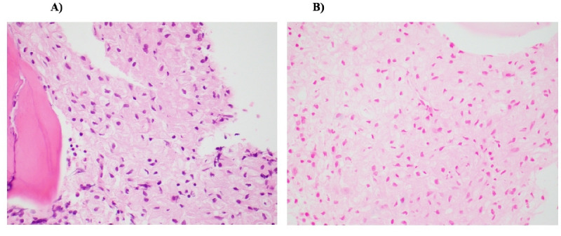
This report presents a case of childhood Gaucher disease type 1, a rare inherited metabolic disorder. Although the clinical symptoms were classical, the histological findings in this case were atypical and initially led to diagnostic uncertainty. The pathognomonic histological finding on bone marrow is Gaucher cells, which are lipid-engorged phagocytes secondary to the accumulation of glucosylceramide. These cells typically demonstrate diffuse and avid iron staining using a Prussian blue iron stain. In this case, although the histiocytes seen on bone marrow were abnormal, the absence of iron staining on bone marrow led to a large range of other diagnoses being considered. In retrospect, this anomaly was likely in the setting of prolonged iron deficiency and anaemia as a result of the insidious nature of this presentation. The prognosis of type 1 Gaucher disease is favourable, with current treatments significantly improving duration and quality of life. We explore the utility of a collaborative multidisciplinary approach in addressing diagnostic uncertainty and the importance in making a diagnosis for Gaucher disease type 1 in order to provide appropriate and targeted treatment.
Keywords: Congenital disorders; Failure to thrive; Paediatrics.
© BMJ Publishing Group Limited 2023. No commercial re-use. See rights and permissions. Published by BMJ.
Conflict of interest statement
Competing interests: None declared.
Figures

References
-
- Tylki-Szymańska A, Vellodi A, El-Beshlawy A, et al. Neuronopathic Gaucher disease: demographic and clinical features of 131 patients enrolled in the International collaborative Gaucher group neurological outcomes Subregistry. J Inherit Metab Dis 2010;33:339–46. 10.1007/s10545-009-9009-6 - DOI - PubMed
Publication types
MeSH terms
Substances
LinkOut - more resources
Full Text Sources
Medical