

Biological properties of experimental dental alginate modified for self-disinfection using green nanotechnology
- PMID: 37775587
- PMCID: PMC10630233
- DOI: 10.1007/s00784-023-05277-8
Biological properties of experimental dental alginate modified for self-disinfection using green nanotechnology
Abstract
Objectives: Disinfection of alginate impression materials is a mandatory step to prevent cross-infection in dental clinics. However, alginate disinfection methods are time-consuming and exert a negative impact on accuracy and mechanical properties. Thus, this study aimed to prepare disinfecting agents (CHX and AgNO3) and silver nanoparticles reduced by a natural plant extract to produce a self-disinfecting dental alginate.
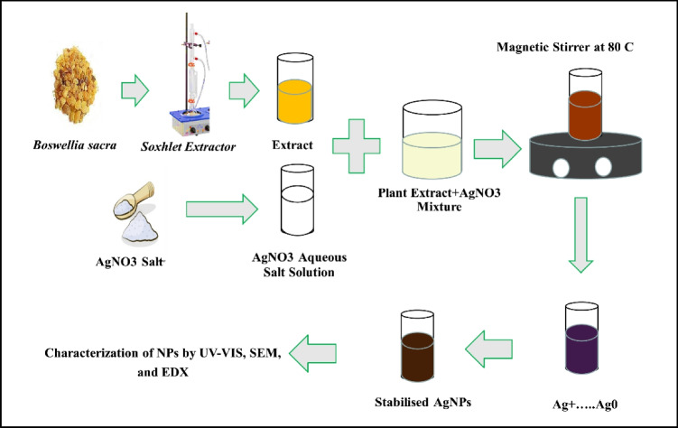
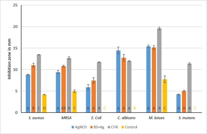
Methods: Conventional alginate impression material was used in this study. Silver nitrate (0.2% AgNO3 group) and chlorohexidine (0.2% CHX group) solutions were prepared using distilled water, and these solutions were later employed for alginate preparation. Moreover, a 90% aqueous plant extract was prepared from Boswellia sacra (BS) oleoresin and used to reduce silver nitrate to form silver nanoparticles that were incorporated in the dental alginate preparation (BS+AgNPs group). The plant extract was characterized by gas chromatography/mass spectrometry (GC/MS) analysis while green-synthesized silver nanoparticles (AgNPs) were characterized by UV-visible (UV-vis) spectroscopy and scanning electron microscopy (SEM). An agar disc diffusion assay was used to test the antimicrobial activity against Candida albicans, Streptococcus mutans, Escherichia coli, methicillin-resistant and susceptible Staphylococcus aureus strains, and Micrococcus luteus. Agar plates were incubated at 37 ± 1 °C for 24 h to allow microbial growth. Diameters of the circular inhibition zones formed around each specimen were measured digitally by using ImageJ software.
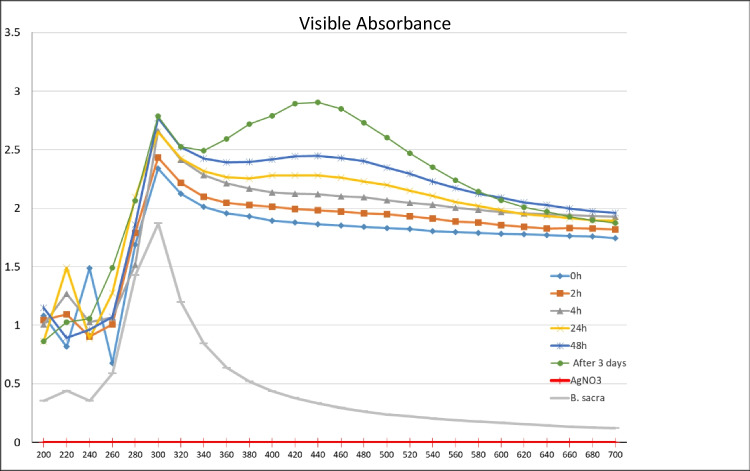
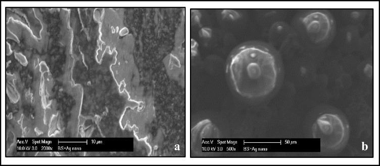
Results: Chemical analysis of the plant extract revealed the presence of 41 volatile and semi-volatile active compounds. UV-Vis spectrophotometry, SEM, and EDX confirmed the formation of spherical silver nanoparticles using the BS extract. CHX, AgNO3, and the BS+AgNPs modified groups showed significantly larger inhibition zones than the control group against all tested strains. BS+AgNPs and CHX groups showed comparable efficacy against all tested strains except for Staphylococcus aureus, where the CHX-modified alginate had a significantly higher effect.
Conclusions and clinical relevance: CHX, silver nitrate, and biosynthesized silver nanoparticles could be promising inexpensive potential candidates for the preparation of a self-disinfecting alginate impression material without affecting its performance. Green synthesis of metal nanoparticles using Boswellia sacra extract could be a very safe, efficient, and nontoxic way with the additional advantage of a synergistic action between metal ions and the phytotherapeutic agents of the plant extract.
Keywords: Antimicrobial activity; Boswellia sacra; Green synthesis; Irreversible hydrocolloids; Nanotechnology.
© 2023. The Author(s).
Conflict of interest statement
All authors declare that there is no conflict of interest in this study.
Figures


Similar articles
-
Mechanical and Physical Properties of an Experimental Chemically and Green-Nano Improved Dental Alginate after Proven Antimicrobial Potentials.Gels. 2023 May 21;9(5):429. doi: 10.3390/gels9050429. Gels. 2023. PMID: 37233020 Free PMC article.
-
Assessing the impact of an environmentally friendly approach on irreversible dental hydrocolloid performance.Sci Rep. 2024 Dec 16;14(1):30516. doi: 10.1038/s41598-024-83035-w. Sci Rep. 2024. PMID: 39681606 Free PMC article.
-
Enhancing Dental Alginate with Syzygium aromaticum, Zingiber officinale and Green Silver Nanoparticles: A Nature-Enhanced Approach for Superior Infection Control.Gels. 2024 Sep 20;10(9):600. doi: 10.3390/gels10090600. Gels. 2024. PMID: 39330202 Free PMC article.
-
Plant Extract-Synthesized Silver Nanoparticles for Application in Dental Therapy.Pharmaceutics. 2022 Feb 8;14(2):380. doi: 10.3390/pharmaceutics14020380. Pharmaceutics. 2022. PMID: 35214112 Free PMC article. Review.
-
Applications of Silver Nanoparticles in Dentistry: Advances and Technological Innovation.Int J Mol Sci. 2021 Mar 2;22(5):2485. doi: 10.3390/ijms22052485. Int J Mol Sci. 2021. PMID: 33801230 Free PMC article. Review.
Cited by
-
Evaluation of Deformation and Antibacterial Properties of Dental Alginates Mixed with Silver Nanoparticles.Materials (Basel). 2025 Apr 30;18(9):2069. doi: 10.3390/ma18092069. Materials (Basel). 2025. PMID: 40363572 Free PMC article.
-
Mechanical and Physical Properties of an Experimental Chemically and Green-Nano Improved Dental Alginate after Proven Antimicrobial Potentials.Gels. 2023 May 21;9(5):429. doi: 10.3390/gels9050429. Gels. 2023. PMID: 37233020 Free PMC article.
-
Assessing the impact of an environmentally friendly approach on irreversible dental hydrocolloid performance.Sci Rep. 2024 Dec 16;14(1):30516. doi: 10.1038/s41598-024-83035-w. Sci Rep. 2024. PMID: 39681606 Free PMC article.
-
Evaluation of the Antibacterial and Antifungal Efficacy of Chitosan Nanoparticles in Irreversible Hydrocolloid Impression Materials: A Cross-Sectional Study.Cureus. 2025 Feb 3;17(2):e78414. doi: 10.7759/cureus.78414. eCollection 2025 Feb. Cureus. 2025. PMID: 40046398 Free PMC article.
-
Antimicrobial Activity of Citrate-Coated Cerium Oxide Nanoparticles.Nanomaterials (Basel). 2024 Feb 13;14(4):354. doi: 10.3390/nano14040354. Nanomaterials (Basel). 2024. PMID: 38392727 Free PMC article.
References
-
- van Noort R. Introduction to dental materials. 3. Mosby, Elsevier; 2007. pp. 191–195.
-
- Saji S, Hebden A, Goswami P, Du C. A Brief review on the development of alginate extraction process and its sustainability. Sustainability. 2022;14:5181. doi: 10.3390/su14095181. - DOI
-
- Shen C, Rawls HR. Phillips’ science of dental materials. 18. St. Louis: Elsevier/Saunders; 2013.
-
- Cervino G, Fiorillo L, Herford AS, Laino L, Troiano G, Amoroso G, Crimi S, Matarese M, D’Amico C, Nastro Siniscalchi E, Cicciù M. Alginate materials and dental impression technique: a current state of the art and application to dental practice. Mar Drugs. 2018;17:18. doi: 10.3390/md17010018. - DOI - PMC - PubMed
MeSH terms
Substances
LinkOut - more resources
Full Text Sources
Molecular Biology Databases
Miscellaneous
