. 2023 Sep 29;14(1):161.
doi: 10.1186/s13244-023-01472-z.

Core and penumbra estimation using deep learning-based AIF in association with clinical measures in computed tomography perfusion (CTP)

Affiliations

Core and penumbra estimation using deep learning-based AIF in association with clinical measures in computed tomography perfusion (CTP)

Sukhdeep Singh Bal et al. Insights Imaging. .

Abstract

Objectives: To investigate whether utilizing a convolutional neural network (CNN)-based arterial input function (AIF) improves the volumetric estimation of core and penumbra in association with clinical measures in stroke patients.

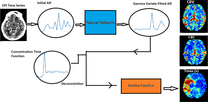
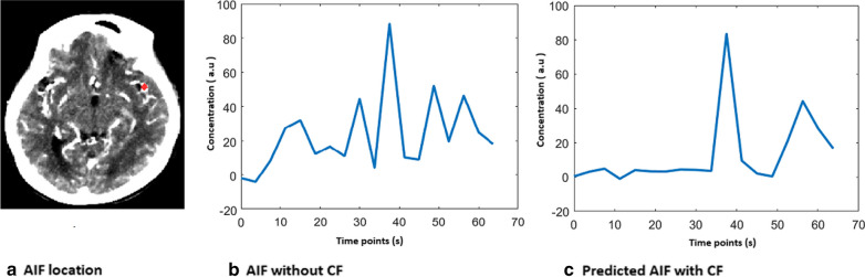
Methods: The study included 160 acute ischemic stroke patients (male = 87, female = 73, median age = 73 years) with approval from the institutional review board. The patients had undergone CTP imaging, NIHSS and ASPECTS grading. convolutional neural network (CNN) model was trained to fit a raw AIF curve to a gamma variate function. CNN AIF was utilized to estimate the core and penumbra volumes which were further validated with clinical scores.

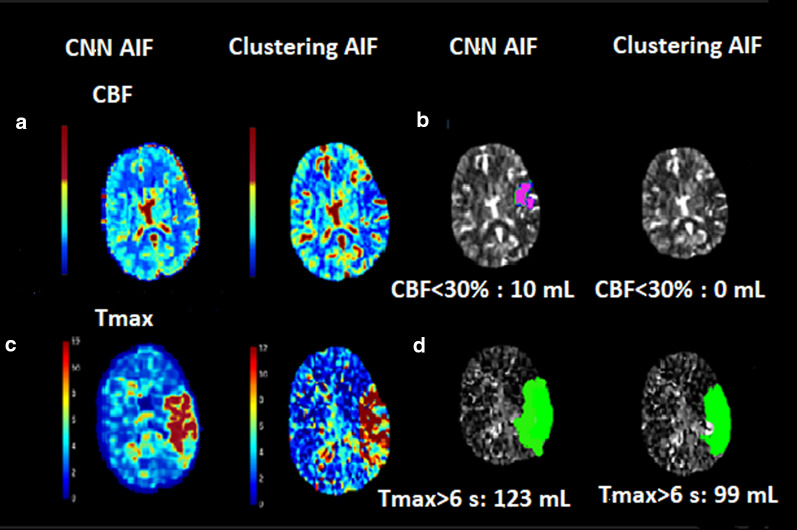
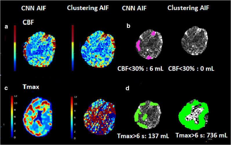
Results: Penumbra estimated by CNN AIF correlated positively with the NIHSS score (r = 0.69; p < 0.001) and negatively with the ASPECTS (r = - 0.43; p < 0.001). The CNN AIF estimated penumbra and core volume matching the patient symptoms, typically in patients with higher NIHSS (> 20) and lower ASPECT score (< 5). In group analysis, the median CBF < 20%, CBF < 30%, rCBF < 38%, Tmax > 10 s, Tmax > 10 s volumes were statistically significantly higher (p < .05).

Conclusions: With inclusion of the CNN AIF in perfusion imaging pipeline, penumbra and core estimations are more reliable as they correlate with scores representing neurological deficits in stroke.

Critical relevance statement: With CNN AIF perfusion imaging pipeline, penumbra and core estimations are more reliable as they correlate with scores representing neurological deficits in stroke.

Keywords: Arterial input function; Core; Ischemic stroke; Penumbra; Perfusion parameters.


Conflict of interest statement

The authors report no competing interest.

Figures

Fig. 1
Fig. 1
Workflow to estimate perfusion parameters
Fig. 2
Fig. 2
Curve fitted AIF for a single dataset (a) AIF location is marked as a red dot. b AIF curve without CF. c Predicted gamma curve fitted AIF curve by CNN model as an output
Fig. 3
Fig. 3
Volumetric agreement of the penumbra (Tmax > 6 s) and infract volume (CBF < 30%) with NIHSS and ASPECTS. a Association of Tmax > 6 s volume with NIHSS. b Association of Tmax > 6 s volume with ASPECTS. c Bland–Altman plot between the penumbra estimated by CNN CF and without CF. d Association of CBF < 30% volume with NIHSS. e Association of CBF < 30% volume with ASPECTS. f Bland–Altman plot between the infract core estimated by CNN CF and without CF
Fig. 4
Fig. 4
Comparison of infarct core and penumbra for a patient with severe stroke. a CBF maps derived with CF CNN AIF and without CF. b Infarct core (CBF < 30% mL) estimated with CF CNN AIF and without CF. c Tmax maps derived with CF and without CF. d Penumbra (Tmax > 6.0 s mL) estimated with CF CNN CF and without CF
Fig. 5
Fig. 5
Comparison of infarct core and penumbra for another patient with severe stroke. a CBF maps derived with CF CNN AIF and without CF. b Infarct core (CBF < 30% mL) estimated with CF CNN AIF and without CF. c Tmax maps derived with CF and without CF. d Penumbra (Tmax > 6.0 s mL) estimated with CF CNN CF and without CF

References

    1. Campbell BCV, Christensen S, Levi CR, et al. Comparison of computed tomography perfusion and magnetic resonance imaging perfusion-diffusion mismatch in ischemic stroke. Stroke. 2012;43:2648–2653. doi: 10.1161/STROKEAHA.112.660548. - DOI - PubMed
    1. Winder A, d’Esterre CD, Menon BK, Fiehler J, Forkert ND. Automatic arterial input function selection in CT and MR perfusion datasets using deep convolutional neural networks. Med Phys. 2020;47:4199–4211. doi: 10.1002/mp.14351. - DOI - PubMed
    1. Suh CH, Jung SC, Cho SJ, et al. Perfusion CT for prediction of hemorrhagic transformation in acute ischemic stroke: a systematic review and meta-analysis. Eur Radiol. 2019;29:4077–4087. doi: 10.1007/s00330-018-5936-7. - DOI - PubMed
    1. Warach S. New imaging strategies for patient selection for thrombolytic and neuroprotective therapies. Neurology. 2001;57:S48–52. doi: 10.1212/WNL.57.suppl_2.S48. - DOI - PubMed
    1. Donnan GA, Davis SM. Breaking the 3 h barrier for treatment of acute ischaemic stroke. Lancet Neurol. 2008;7:981–982. doi: 10.1016/S1474-4422(08)70230-8. - DOI - PubMed

LinkOut - more resources