. 2023 Sep 29;13(1):16420.
doi: 10.1038/s41598-023-41942-4.

New drug discovery of cardiac anti-arrhythmic drugs: insights in animal models

Affiliations

New drug discovery of cardiac anti-arrhythmic drugs: insights in animal models

Ashish Kumar Sharma et al. Sci Rep. .

Abstract

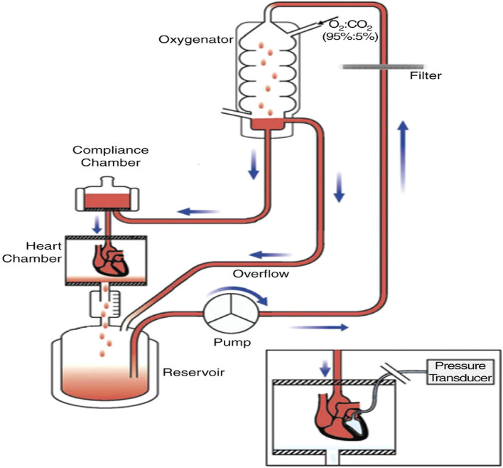
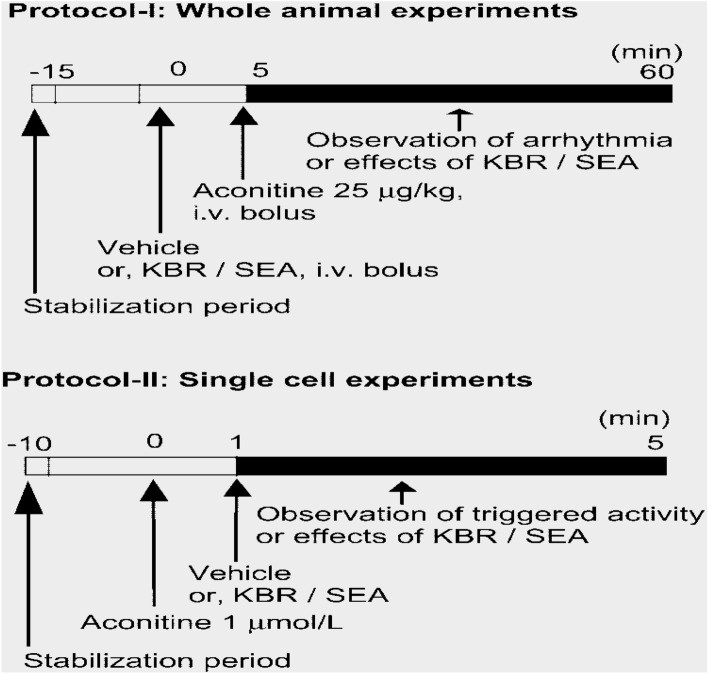
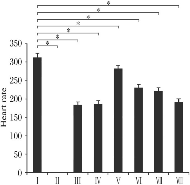
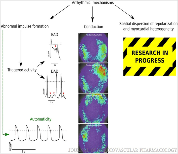
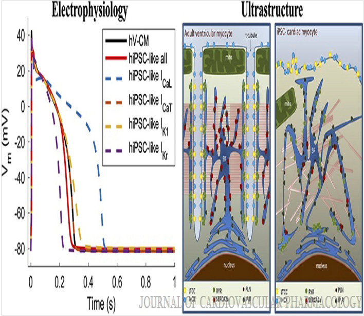
Cardiac rhythm regulated by micro-macroscopic structures of heart. Pacemaker abnormalities or disruptions in electrical conduction, lead to arrhythmic disorders may be benign, typical, threatening, ultimately fatal, occurs in clinical practice, patients on digitalis, anaesthesia or acute myocardial infarction. Both traditional and genetic animal models are: In-vitro: Isolated ventricular Myocytes, Guinea pig papillary muscles, Patch-Clamp Experiments, Porcine Atrial Myocytes, Guinea pig ventricular myocytes, Guinea pig papillary muscle: action potential and refractory period, Langendorff technique, Arrhythmia by acetylcholine or potassium. Acquired arrhythmia disorders: Transverse Aortic Constriction, Myocardial Ischemia, Complete Heart Block and AV Node Ablation, Chronic Tachypacing, Inflammation, Metabolic and Drug-Induced Arrhythmia. In-Vivo: Chemically induced arrhythmia: Aconitine antagonism, Digoxin-induced arrhythmia, Strophanthin/ouabain-induced arrhythmia, Adrenaline-induced arrhythmia, and Calcium-induced arrhythmia. Electrically induced arrhythmia: Ventricular fibrillation electrical threshold, Arrhythmia through programmed electrical stimulation, sudden coronary death in dogs, Exercise ventricular fibrillation. Genetic Arrhythmia: Channelopathies, Calcium Release Deficiency Syndrome, Long QT Syndrome, Short QT Syndrome, Brugada Syndrome. Genetic with Structural Heart Disease: Arrhythmogenic Right Ventricular Cardiomyopathy/Dysplasia, Dilated Cardiomyopathy, Hypertrophic Cardiomyopathy, Atrial Fibrillation, Sick Sinus Syndrome, Atrioventricular Block, Preexcitation Syndrome. Arrhythmia in Pluripotent Stem Cell Cardiomyocytes. Conclusion: Both traditional and genetic, experimental models of cardiac arrhythmias' characteristics and significance help in development of new antiarrhythmic drugs.


Conflict of interest statement

The authors declare no competing interests.

Figures

Figure 1
Figure 1
Schematic of the cardiac conduction system and clinical classification of cardiac arrhythmias. AF indicates atrial fibrillation; AV, atrioventricular; AVNRT, AV nodal reentry tachycardia; AVRT, AV reciprocating tachycardia; PAC, premature atrial; and SA, sinoatrial. Illustration credit: Ben Smith. [Daniel J. Blackwell. Circulation Research. Animal Models to Study Cardiac Arrhythmias, Volume: 130, Issue: 12, Pages: 1926–1964, (10.1161/CIRCRESAHA.122.320258)].
Figure 2
Figure 2
The ventricular action potential and ionic currents in humans and mice. Note the differences in action potential shape, which is caused primarily by differences in ionic currents circled in red. ICa(L) indicates L-type Ca current; ICa(T), T-type Ca current; INa, Na current; INaCa, Na-Ca-Exchange current; INaK, NaK-ATPase pump current; IK1, Inward rectifier K-current; IKr, rapidly activating delayed rectifier K-current; IKs, slowly activating delayed rectifier K-current; Iss, rapidly activating steady-state K-currents; and Ito, transient outward K-current. Please note that current densities (pA/pF) measured in single cells vary drastically with experimental conditions and voltage clamp protocols. Current densities were chosen to reflect relative contributions to the AP. [Daniel J. Blackwell. Circulation Research. Animal Models to Study Cardiac Arrhythmias, Volume: 130, Issue: 12, Pages: 1926–1964 (10.1161/CIRCRESAHA.122.320258)].
Figure 3
Figure 3
Tissue-targeted CASQ2 knock-out mice help decipher the anatomic origin of ventricular ectopy in catecholaminergic polymorphic ventricular tachycardia (CPVT). Based on a combination of tissue-targeting and in silico modeling, the PMJ was identified as the likely origin of ventricular ectopy in CPVT. DAD indicates delayed afterdepolarization; LV, left ventricular; PMJ, Purkinje myocardial junction; and RV, right ventricular. Illustration credit: Ben Smith. [Daniel J. Blackwell. Circulation Research. Animal Models to Study Cardiac Arrhythmias, Volume: 130, Issue: 12, Pages: 1926–1964 (10.1161/CIRCRESAHA.122.320258)].
Figure 4
Figure 4
Prepration of rabbit auricles suspended in oxygenated Ringer-Locke at 29 °C. The electrode holder is made of perpex, and the inset shows a closer view of its lower end. (Dawes GS. Synthetic substitutes for quinidine. British Journal of Pharmacology and Chemotherapy. 1946 Jun;1(2):90–112. PMCID: PMC1509732 PMID: 19108083).
Figure 5
Figure 5
Shows the experimental methodology and the separated core. (A). Isolated guinea pig heart installed on the Langendorff apparatus showing the front view with all the electrodes and probes in place. (B). The usual procedure required at least 30 min for cardiac perfusion stabilization, followed by four periods of 15 min each for exposure to baseline and vehicle in three escalating doses.CPP, LVP, ECG and MAP were continuously monitored and recorded throughout the study. The horizontal bars show that the heart is beating freely and automatically, except slowly. (Guo L, Dong Z, Guthrie H. Validation of a guinea pig Langendorff heart model for assessing potential cardiovascular liability of drug candidates. Journal of Pharmacological and Toxicological Methods. 2009 Sep-Oct, 60(2):130–151. PMID: 19616638. 10.1016/j.vasch.2009.07.002)).
Figure 6
Figure 6
Isolated Langendorff heart perfusion model, (Ravelli F, Allessie MA. Effects of atrial dilatation on refractory period and vulnerability to atrial fibrillation in the isolated Langendorff-perfused rabbit heart. Circulation 1997 Sep 2;96(5):1686–95. PMID: 9315565. 10.1161/01.cir.96.5.1686)).
Figure 7
Figure 7
Overview of the test protocols. Protocol I was used for whole animal experiments and Protocol II for single cell experiments. In Protocol I, after stabilization, the medium or each dose of KBR or SEA was pretreated. Then aconitine (25 g/kg, IV)bolus has been delivered). The black column indicates the duration of the arrhythmia observation. A total of 130 animals were introduced into this series of experiments. 74 were used in Protocol I, but six were excluded based on exclusion criteria. The number of animals assigned to each control (vehicle), KBR and SEA is shown in Fig. 3. In Protocol II, after stabilization, aconitine (1 M) was infused first, then vehicle, or each dose of KBR or SEA was further elaborated. A total of 13 cells were included in Protocol II(Amran MS, Hashimoto K, Homma N. Effects of sodium-calcium exchange inhibitors, KB-R7943 and SEA0400, on aconitine-induced arrhythmias in guinea pigs in vivo, in vitro, and in computer simulation studies. Journal of Pharmacology and Experimental Therapeutics. 2004 Jul 1;310(1):83–9. PMID: 15028781. 10.1124/jpet.104.066951) ).
Figure 8
Figure 8
A sample ECG for Group II (10% CaCl2) taken at various times. In Group II [Arrhythmia control group 1, 10% CaCl2 (50 mg/kg) (n = 6)], the ECGs were found. Using 10% CaCl2 dose (at various times after intravenous injection). After administering 10% CaCl2, arrhythmia, cardiodepression, and death were noted at 20 min. (A) Normal reading (Control); (B) Arrhythmia caused by 10% CaCl2 dose at 0 min after administration; (C) Arrhythmia caused by 10% CaCl2 dose at 5 min after administration; (D) Arrhythmia caused by 10% CaCl2 dose at 10 min after administration; (E) Complete cardiodepression caused by 10% CaCl2 dose at 15 min after administration; (F) Mortality caused by 10% CaCl2 dose at 20 min after administration. . (Sharma AK, Kishore K, Sharma D, Srinivasan BP, Agarwal SS, Sharma A, Singh SK, Gaur S, Jatav VS. Cardioprotective activity of alcoholic extract of Tinospora cordifolia (Wild.) Miers in calcium chloride-induced cardiac arrhythmia in rats. Journal of Biomedical Research. 2011 Jul 1;25(4):280–6. PMID: 23554702. 10.1016/S1674-8301(11)60038-9)).
Figure 9
Figure 9
Shows an ECG that is representative of the several groups (Group I to Group VIII, n = 6) (A) Standard reading (Control); (B) Group III: "5% CaCl2 (25 mg/kg)" for arrhythmia management; (C) "CaCl2 (5%)-induced arrhythmia + alcohol (95%, 0.5 ml, IV); (D) Group V: Verapamil (5 mg/kg, IV) with CaCl2 (5%)-induced arrhythmia; (E) Tinospora cordifolia dried ethanolic extract dissolved in saline plus Group VI CaCl2 (5%)-induced arrhythmia.Tinospora cordifolia dried ethanolic extract diluted in saline (150 mg/kg, iv); Group VII CaCl2 (5%)-induced arrhythmia + Tinospora cordifolia extract (250 mg/kg, iv); Group VIII CaCl2 (5%)-induced arrhythmia + Tinospora cordifolia dry ethanolic extract (450 mg/kg, iv) (n = 6). ). (Sharma AK, Kishore K, Sharma D, Srinivasan BP, Agarwal SS, Sharma A, Singh SK, Gaur S, Jatav VS. Cardioprotective activity of alcoholic extract of Tinospora cordifolia (Wild.) Miers in calcium chloride-induced cardiac arrhythmia in rats. Journal of Biomedical Research. 2011 Jul 1;25(4):280–6. PMID: 23554702. 10.1016/S1674-8301(11)60038-9) ).
Figure 10
Figure 10
Shows a comparison of the effects of verapamil and Tinospora cordifolia extract on heart rate. *P 0.05 indicates statistical significance compared to control (Group I). (n = 6). CaCl2 (5%) + verapamil (5 mg/kg, iv); Group VI: CaCl2 (5%) + Tinospora cordifolia (150 mg/kg, iv); Group VII: CaCl2 (5%) + Tinospora cordifolia (250 mg/kg, iv); Group VIII: CaCl2 (5%) + Tinospora cordifolia (450 mg/kg, iv).Group I: control. (Sharma AK, Kishore K, Sharma D, Srinivasan BP, Agarwal SS, Sharma A, Singh SK, Gaur S, Jatav VS. Cardioprotective activity of alcoholic extract of Tinospora cordifolia (Wild.) Miers in calcium chloride-induced cardiac arrhythmia in rats. Journal of Biomedical Research. 2011 Jul 1;25(4):280–6. PMID: 23554702. 10.1016/S1674-8301(11)60038-9)).
Figure 11
Figure 11
Scope of the review. Although arrhythmogenic properties of hiPSC-CMs are very relevant in all 3 principal application fields (basic research, drug screening, and regenerative therapy), we focus on the first one and on the second one to a lesser extent. Also, we link the hiPSC-based findings to the human/patient context whenever possible. (Paci, Michelangelo; Penttinen, Kirsi; Pekkanen-Mattila, Mari; Koivumäki, Jussi T. Arrhythmia Mechanisms in Human Induced Pluripotent Stem Cell–Derived Cardiomyocytes. Journal of Cardiovascular Pharmacology77(3):300–316, March 2021. 10.1097/FJC.0000000000000972).
Figure 12
Figure 12
The classification of arrhythmic mechanisms used in this review article. The panels for EADs and DADs were adapted from Ref. 43 under CC BY 3.0 (original Figs. 5A and 7G, http://creativecommons.org/licenses/by/3.0/). The panel for automaticity was adapted from Ref. 142 under CC BY 3.0 (original Fig. 2A). The panel for conduction was adapted from Ref. 77 under CC BY 4.0 (original Supplementary Movie 5, http://creativecommons.org/licenses/by/4.0/). (Paci, Michelangelo; Penttinen, Kirsi; Pekkanen-Mattila, Mari; Koivumäki, Jussi T. Arrhythmia Mechanisms in Human Induced Pluripotent Stem Cell–Derived Cardiomyocytes. Journal of Cardiovascular Pharmacology 77(3):300–316, March 2021. 10.1097/FJC.0000000000000972).
Figure 13
Figure 13
Distinct electrophysiological and ultrastructural characteristics of hiPSC-CMs in comparison with human native/adult ventricular CMs (hV-CMs). Impact of prototypical hiPSC-like ICaL, IK1, and IKr densities, which are 1.5-fold, 0.5-fold, and fourfold in comparison with hV-CM, on the repolarization reserve, simulated as in Ref. 193. The absence of t-tubules in hiPSC-CM results in spatial uncoupling of L-type Ca2 + channel (LTCC) and ryanodine receptors (RyR), as well as irregular distribution of SR Ca2 + handling proteins: RyR, Ca2 + ATPase (SERCA), and its regulatory protein phospholamban (PLN). Furthermore, inositol triphosphate receptors (IP3R) activity is substantially higher in hiPSC-CMs. The panels for were adapted from Ref. 196 CC BY 4.0 (original Fig. 1, http://creativecommons.org/licenses/by/4.0/). (Paci, Michelangelo; Penttinen, Kirsi; Pekkanen-Mattila, Mari; Koivumäki, Jussi T. Arrhythmia Mechanisms in Human Induced Pluripotent Stem Cell–Derived Cardiomyocytes. Journal of Cardiovascular Pharmacology77(3):300–316, March 2021. 10.1097/FJC.0000000000000972).

References

    1. Roden DM. Antiarrhythmie Drugs. In: Laurence LB, Lazo JS, Parker KL, editors. Goodman & Gilman's The Pharmcological Basis of Thaerapeutics. 11. McGraw Hill; 2006. pp. 899–932.
    1. Khurshid S, Choi SH, Weng LC, Wang EY, Trinquart L, Benjamin EJ, Ellinor PT, Lubitz SA. Frequency of cardiac rhythm abnormalities in a half million adults. Circ. Arrhythm. Electrophysiol. 2018;11:e006273. doi: 10.1161/CIRCEP.118.006273. - DOI - PMC - PubMed
    1. Echt DS, Liebson PR, Mitchell LB, Peters RW, Obias-Manno D, Barker AH, Arensberg D, Baker A, Friedman L, Greene HL. Mortality and morbidity in patients receiving encainide, flecainide, or placebo. The cardiac arrhythmia suppression trial. N. Engl. J. Med. 1991;324:781–788. doi: 10.1056/NEJM199103213241201. - DOI - PubMed
    1. Cardiac Arrhythmia Suppression Trial III Effect of the antiarrhythmic agent moricizine on survival after myocardial infarction. N. Engl. J. Med. 1992;327:227–233. doi: 10.1056/NEJM199207233270403. - DOI - PubMed
    1. Delisle BP, Anson BD, Rajamani S, January CT. Biology of cardiac arrhythmias: Ion channel protein trafficking. Circ Res. 2004;94:1418–1428. doi: 10.1161/01.RES.0000128561.28701.ea. - DOI - PubMed